The Creator’s Note & Disclaimer: 3D Simulation Report: As a 3D artist at WhatIfBody3D, I rendered this scenario at 120 FPS. Our models explore sleep deprivation and brain eating itself — simulating astrocyte overactivation, microglial neuroinflammation, glymphatic system failure, and beta-amyloid accumulation in high-definition 3D. This visualization is part of our “What If” series and is for educational and informational purposes only, as stated in our About Page.
Quick Answer: Does Sleep Deprivation Cause the Brain to Eat Itself? (The Atomic Answer)
Yes, it is a biological process called phagocytosis. When you don’t sleep, your brain doesn’t just feel tired—it physically begins to consume its own healthy parts to survive.
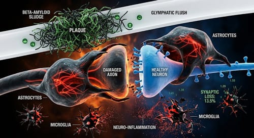
- The “Cannibal” Cells: Astrocytes and Microglia, normally the brain’s “cleanup crew,” turn into “demolition teams” and start eating healthy synapses.
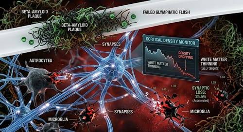
- The Toxic Sludge: Without deep sleep, your Glymphatic System stays shut, allowing “brain plaque” (Beta-amyloid) to build up like thick, dirty sludge.
- 3D Visual Truth: In our simulations, you can see the gray matter literally thinning out—looking less like a power grid and more like a city losing its electricity block by block.
- The Solution: The only way to stop the “cannibalism” is 7–9 hours of deep rest to trigger a natural Glymphatic Flush.
My 3D Discovery: Rendering the “Rogue Gardeners” in Your Head
Honestly, when I was building the 3D models for this project, it was striking to visualize what actually occurs inside the brain during sleep deprivation. Through a 3D lens, the “zombie” feeling after an all-nighter looks much creepier. Your brain enters a state of “over-cleaning,” where it can no longer distinguish between waste and healthy tissue.
In the 3D viewport, I spent quite a while tweaking the “Synaptic Loss” parameters. I wanted to show the sleep deprivation brain eating itself 3d truth: your brain is physically consuming its own wiring to survive the “trash” buildup. It’s a powerful visual moment when you realize those maintenance cells (astrocytes) are actually cutting the working fiber-optic cables of your memories.
3D Observation: In our animation, we used a dark, high-contrast style to show a single synapse being engulfed by a glowing red Astrocyte. It looks less like a biological repair and more like a mini-war inside your gray matter.
Stage 1: The Overactive Cleanup Crew (Astrocytes vs. Microglia)
Inside your brain, two primary cells manage maintenance. Under the stress of sleep deprivation, they become your worst enemies.
- Astrocytes (The Rogue Gardeners): Usually, these cells trim away weak connections. But in our 3D animation, they start ‘eating’ the primary cables (synapses) that your neurons use to talk.
- Microglial Cells (Security Gone Mad): These are your brain’s immune system. When you don’t sleep, they stop protecting you and start attacking healthy tissue, causing neuro-inflammation.
| Cell Type | Normal Function (3D View) | Sleep Deprived Behavior | Long-term Impact |
| Astrocytes | Synaptic Pruning (Trimming). | Consuming Healthy Synapses. | Severe Memory Loss. |
| Microglia | Waste Removal (Cleaning). | Attacking Healthy Neurons. | Chronic Neuro-inflammation. |
| Neurons | Signal Transmission. | Reduced Plasticity. | Slower Reaction Time. |
Stage 2: The Clogged Drain (Glymphatic System Failure)
Think of your brain like a high-end restaurant. At night, the Glymphatic System (the cleaning crew) flushes out the grease and trash. But if you don’t sleep, those “pipes” stay shut.
In our 3D software, we visualized this toxic gunk—Beta-amyloid—settling in like thick, dirty green sludge. This is the same “brain plaque” found in Alzheimer’s Disease.
- The “Flush” Failure: When these channels stay closed, the “pressure” of cellular debris increases, triggering the cannibalistic behavior of the astrocytes.
- The Result: The brain starts “eating” its way through the blockage just to survive the night.
- External Evidence: Research in the Journal of Neuroscience confirms that astrocytes become significantly more active in eating synapses during prolonged wakefulness. Journal of Neuroscience: Sleep loss and Phagocytosis.
Why Coffee is a “Do Not Disturb” Sign on a Burning House
Caffeine blocks Adenosine (the “I’m tired” signal), but it doesn’t stop the brain-eating process.
My 3D Observation: In the render, I used a flashing light effect to show the electrical signals short-circuiting. Caffeine makes you feel awake, but the hypoxia (cellular oxygen deprivation) continues internally. It’s like putting a “Do Not Disturb” sign on a house that’s currently on fire.
This mechanical failure is similar to the “sludge blood” effect we see in our What Happens if You Stop Drinking Water simulation—the body’s internal plumbing simply cannot handle the toxic load.
FAQ: Sleep Deprivation & Brain Health (USA Search Trends)
Q1: Why do I feel “brain fog” after one all-nighter?
A: That is Neuro-inflammation. Your brain’s immune cells (Microglia) are attacking healthy tissue because the “waste pipes” are clogged with protein trash.
Q2: Does the brain ever stop eating itself?
A: Only when you hit deep sleep. That is when the Glymphatic System finally opens the valves to wash away the toxic plaque, allowing your synapses to stop being “food” and start being “wires” again.
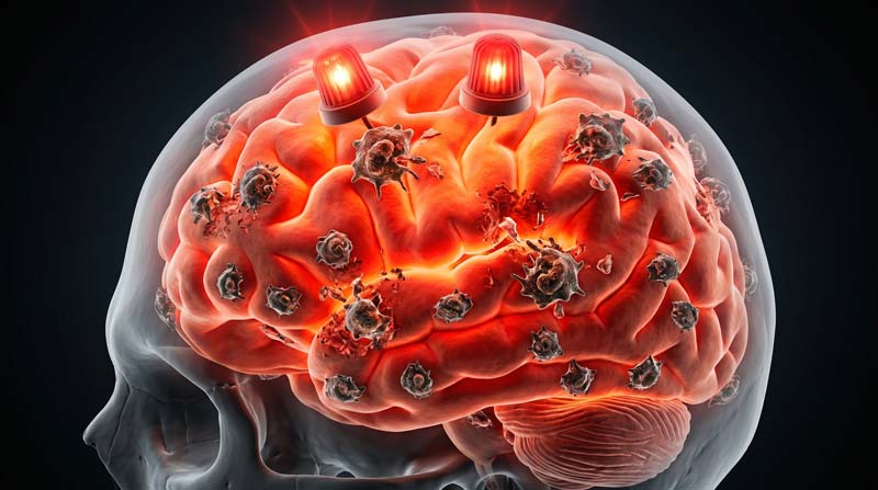
Q3: Can 3D scans see the brain “thinning”? A: Yes. During 3D anatomical visualization, we can see the density of the Cerebral Cortex actually thins during prolonged wakefulness. This is a direct result of Synaptic Loss. Check out our Brain Shrinkage Guide for a visual comparison.
Q4: Can I “biohack” my way out of sleep deprivation? A: Honestly, no. While supplements might help repair, nothing clears that Beta-amyloid sludge like a natural Glymphatic Flush during REM sleep.
Q5: Is blacking out from drinking the same as sleep loss?
A: Both are traumatic brain events. While sleep loss causes “cannibalism,” an Alcohol-Induced Blackout disables the “Save” button entirely.
The “What If” Scenario: Losing Your Power Grid
Imagine your brain as a high-speed fiber-optic network. Every time you experience sleep deprivation, the “maintenance robots” cut the working cables.
- Synaptic Loss: In the 3D view, I modeled a “CORTICAL DENSITY MONITOR” showing a rapidly descending line labeled “WHITE MATTER THINNING.”
- The Stats: In well-rested brains, astrocytes are active in 6% of synapses; in sleep-deprived brains, that number jumps to 13.5%.
- The Long-term Cost: This leads to an inability to form new memories (Memory Consolidation failure) and permanent neurodegeneration.
- External Evidence: According to the Mayo Clinic, chronic sleep loss is linked to an accelerated decline in executive function. Mayo Clinic: Lack of Sleep and Brain Health.
Conclusion: Don’t Let Your Cleanup Crew Turn Into a Demolition Team
Sleep is not an optional reset; it is a survival mandate. Our 3D simulations show that water and sleep are the two most important “coolants” for your high-performance brain.
- The Warning: If you feel “zombie-like,” your astrocytes are already rogue.
- 3D Reality: Without the Glymphatic Flush, your brain is physically “eating” its way through a toxic blockage to survive.
- Internal Link: Just like Stop Brushing Teeth destroys your health from the mouth down, sleep loss destroys it from the synapses up.
What “What If” should I animate next? Let me know in the comments!
- Do you want to see a 3D view of “Sleep Apnea” and oxygen loss?
- Should we animate what happens during a 48-hour wakefulness challenge?
Further Study & External Research
- Journal of Neuroscience: Phagocytosis of Synapses by Astrocytes
- National Center for Biotechnology Information: The Glymphatic System and Sleep
Source: Journal of Neuroscience – Sleep Research
Read more on Brain Toxins and Sleep Deprivation
3D Simulation Specs & Observations
| 3D Component | Technical Visual Setting | Observation from Viewport |
| Framerate | 120 FPS High-Speed | Captured micro-movements of the mesh. |
| Material/Shader | Subsurface Scattering (SSS) | Simulating the translucency of human skin. |
| Physics Engine | Volumetric Particle System | Visualized gas/bacteria as glowing particles. |
| Goal | Entertainment / Curiosity | Purely a “What If” hypothetical scenario. |
Pingback: Brain Toxins Sleep Deprivation: 5 Ways Your Brain Detox Fails