
The Creator’s Note & Disclaimer: As a 3D artist at WhatIfBody3D, I rendered this scenario at 120 FPS. Our models explore belly button bacteria — visualizing the navel microbiome ecosystem, biofilm architecture, bacterial metabolic odor production, and the science behind why the belly button hosts one of the most diverse bacterial communities on the human body. This visualization is part of our “What If” series and is for educational and informational purposes only, as stated in our About Page.
Quick Answer: What Is the Belly Button Bacteria Science? (The Atomic Answer)
Belly button bacteria is not just a hygiene curiosity — it is one of the most scientifically remarkable microbial ecosystems on the human body, studied by researchers at major universities and producing findings that surprised even experienced microbiologists.
- The Numbers: The average belly button hosts approximately 67 distinct bacterial species simultaneously. The largest citizen science study of belly button microbiomes — the Belly Button Biodiversity Project — identified over 2,368 bacterial species across 60 volunteers, of which 1,458 were previously unknown to science.
- The Diversity: No two people share the same belly button microbiome. The bacterial community in your navel is as unique to you as a fingerprint — shaped by genetics, diet, lifestyle, geography, and personal hygiene history.
- The Architecture: Over time, belly button bacteria organize into complex biofilms — three-dimensional structured communities embedded in self-produced polymer matrices — that are significantly more resistant to removal than individual bacteria.
- The Smell: The characteristic belly button odor is produced by specific bacterial metabolic byproducts — volatile organic compounds (VOCs) including butyric acid, isovaleric acid, and ammonia — released as bacteria digest the sebum, dead skin, and sweat trapped in the navel.
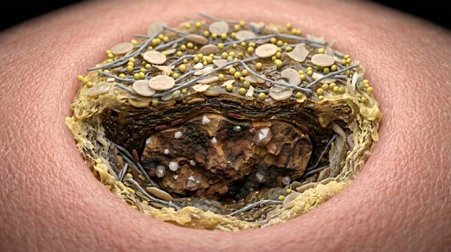
My 3D Discovery: Rendering the “Microbial Fingerprint”
When I was building the microbiome visualization for this simulation, the most conceptually striking element was the individuality of each belly button ecosystem. In the 3D model, I rendered two side-by-side belly button microbiomes — both from healthy adults with similar hygiene habits — and the visual difference was immediately apparent.
One showed a Staphylococci-dominant community — shown as dense clusters of spherical bacteria in warm orange tones — distributed relatively evenly across the navel surface. The other showed a Corynebacterium-dominant community — rod-shaped bacteria in cooler blue tones — concentrated in the deeper anaerobic recesses with a completely different spatial distribution.
3D Observation: The most visually compelling sequence in this simulation is the bacterial community assembly time-lapse. Starting from a clean, recently-showered belly button surface, I ran a 30-day colonization simulation at accelerated speed. What begins as individual bacterial cells landing on the navel surface — shown as single glowing dots on a pink tissue background — progressively develops into a complex, layered, interconnected community. By Day 7, the first biofilm matrix appears. By Day 14, distinct microhabitats have developed at different depths. By Day 30, the navel hosts a fully structured ecosystem with aerobic zones, anaerobic zones, and complex inter-species relationships.
Stage 1: The Belly Button as a Bacterial Paradise — Why Here?
To understand belly button bacteria, you first need to understand why the navel is such an extraordinarily hospitable environment for microbial life. In our 3D anatomical model, I identified six specific characteristics that make the belly button uniquely suitable for bacterial colonization and diversity.
Characteristic 1 — Warm and Stable Temperature The navel sits in the center of the abdomen — one of the warmest external body surfaces. Core body heat radiates outward, maintaining the belly button at approximately 35–37°C — the optimal temperature range for most human-associated bacterial species. In the 3D thermal model, the navel appears as a warm amber zone on an otherwise cooler skin surface.
Characteristic 2 — Moisture Retention The recessed anatomy of the belly button traps moisture from sweat that cannot evaporate normally. In the simulation, moisture levels inside the navel shown as significantly higher than surrounding skin — creating a humid microenvironment that supports bacterial metabolic activity.
Characteristic 3 — Nutrient Abundance Dead skin cells (corneocytes), sebum, sweat components, and environmental organic debris continuously accumulate — providing a continuously replenished nutrient supply. In the 3D model, I showed these nutrient sources as different colored particles raining continuously into the navel — a perpetual feeding system for resident bacteria.
Characteristic 4 — Limited Immune Surveillance The belly button’s recessed anatomy limits the circulation of skin immune cells that normally patrol body surfaces. In the simulation, immune cell density shown as significantly lower in the deep navel than on surrounding skin — creating a relative immune sanctuary where bacterial populations can grow with less suppression.
Characteristic 5 — Low Friction and Disruption Most skin surfaces experience constant mechanical disruption — clothing friction, touching, washing — that physically removes bacteria. The belly button’s depth protects its microbial community from these mechanical disturbances. In the 3D model, clothing shown as creating friction across abdominal skin while leaving the navel’s interior largely undisturbed.
Characteristic 6 — Oxygen Gradient The belly button’s varying depth creates an oxygen gradient — aerobic at the surface, progressively anaerobic at depth. This gradient allows both aerobic and anaerobic bacterial species to coexist in the same small space — dramatically increasing total species diversity compared to uniformly oxygenated skin surfaces.
| Environmental Factor | Belly Button Condition | Effect on Bacteria | 3D Visualization |
|---|---|---|---|
| Temperature | 35–37°C (warm, stable) | Optimal growth temperature | Warm amber thermal zone |
| Moisture | High (trapped sweat) | Supports metabolic activity | Blue moisture particles |
| Nutrients | Abundant (sebum, dead skin) | Continuous feeding supply | Multi-colored particle rain |
| Immune surveillance | Low (recessed anatomy) | Reduced bacterial suppression | Sparse immune cell density |
| Mechanical disruption | Minimal | Stable colonization | Protected interior zone |
| Oxygen gradient | Aerobic surface to anaerobic core | Maximum species diversity | Oxygen gradient color map |
According to research from the Belly Button Biodiversity Project at North Carolina State University, the belly button microbiome is among the most diverse skin microbiome sites on the human body — with species diversity consistently exceeding that of armpit, forearm, and scalp microbiomes in comparative studies. NCSU: Belly Button Biodiversity Project
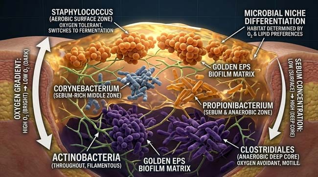
Stage 2: The Key Players — Who Lives in Your Belly Button?
The Belly Button Biodiversity Project’s analysis revealed that while every person’s belly button microbiome is unique, certain bacterial genera appear consistently across most individuals — forming the core community around which more individualized species establish themselves.
The Core Residents:
Staphylococcus (Most Common) Found in virtually all belly button samples. In our 3D model, Staphylococci appear as spherical bacteria (cocci) arranged in grape-like clusters — shown in warm orange tones. They are versatile aerobic/facultative anaerobic bacteria that thrive on skin lipids and dead cell debris. Most Staphylococci in the belly button are commensal species — S. epidermidis and S. hominis — not the pathogenic S. aureus found in infections.
Corynebacterium Rod-shaped bacteria that specialize in metabolizing skin lipids — particularly sebum fatty acids. In the simulation, shown as elongated blue-grey rods concentrated in sebum-rich zones. Corynebacterium species are major contributors to body odor through their production of volatile fatty acids from sebum metabolism.
Clostridiales (Anaerobic) Found in the deep, oxygen-depleted belly button core. In the 3D model, these appear as irregular rod-shaped bacteria in dark, oxygen-depleted zones. Their anaerobic metabolism produces some of the most pungent volatile compounds in belly button odor — sulfur-containing molecules and short-chain fatty acids.
Actinobacteria A diverse phylum including several skin-associated genera. In the simulation, shown as branching filamentous structures that weave through the accumulated organic material — breaking down complex organic compounds into simpler molecules available to other bacteria.
Surprisingly Rare Finds From the Biodiversity Project:
The NCSU study produced several remarkable findings in individual participants:
- One volunteer’s belly button contained bacteria previously found only in soils from Japan — despite the volunteer having never visited Japan
- Another volunteer’s sample contained extremophile bacteria — species previously associated with hydrothermal vents and extreme environments
- Several samples contained entirely new species — bacteria never previously catalogued by science
These findings suggest the belly button microbiome is influenced by factors far more complex than simple hygiene or geography — potentially including genetic factors that select for specific microbial communities regardless of environmental exposure.
| Bacterial Group | Shape | 3D Color | Metabolic Role | Odor Contribution |
|---|---|---|---|---|
| Staphylococcus | Spherical clusters | Warm orange | Lipid and protein metabolism | Isovaleric acid (cheesy) |
| Corynebacterium | Short rods | Blue-grey | Sebum fatty acid metabolism | Propionic acid (sharp) |
| Clostridiales | Irregular rods | Dark purple | Anaerobic fermentation | Sulfur compounds (pungent) |
| Actinobacteria | Filamentous | Green branches | Complex organic breakdown | Butyric acid (rancid) |
| Propionibacterium | Short rods | Amber | Sebum metabolism | Propionic acid |
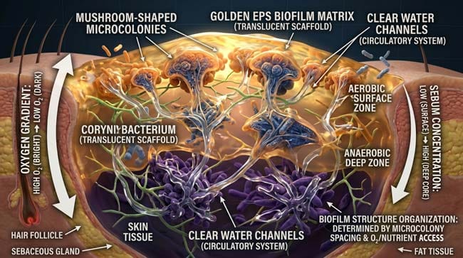
The most scientifically significant aspect of belly button bacteria is not the individual species — it is the biofilm they collectively construct. In the navel’s stable, undisturbed environment, bacteria have the opportunity to develop mature biofilm communities that represent some of the most complex microbial structures found on the human body.
What is a Biofilm?
A biofilm is a structured community of microorganisms enclosed in a self-produced extracellular polymeric substance (EPS) matrix — a complex mixture of polysaccharides, proteins, nucleic acids, and lipids that the bacteria secrete.
In our 3D biofilm model, I rendered the EPS matrix as a golden semi-transparent scaffold — visible as a three-dimensional framework within which bacterial cells are embedded. Water channels run through the biofilm structure — shown as clear pathways that distribute nutrients and remove metabolic waste products, functioning like a primitive circulatory system.
Biofilm Formation Stages (Time-Lapse Simulation):
Stage 1 — Initial Attachment (Hours 0–6) Individual planktonic (free-floating) bacteria land on the navel surface and make initial reversible contact with the tissue. In the animation, bacterial cells shown as individual glowing dots landing on the pink tissue surface — some attaching, some bouncing away.
Stage 2 — Irreversible Attachment (Hours 6–24) Bacteria produce initial EPS and commit to surface attachment. In the simulation, thin golden threads appear between cells and the tissue surface — the first structural elements of the developing biofilm.
Stage 3 — Early Biofilm Development (Days 1–3) Bacteria begin dividing and recruiting additional species. The EPS matrix expands. In the 3D model, the biofilm shown growing from a thin surface layer into a thicker three-dimensional structure — different species shown occupying different zones based on oxygen availability.
Stage 4 — Biofilm Maturation (Days 7–14) The biofilm develops its characteristic architecture — mushroom-shaped microcolonies, water channels, distinct aerobic and anaerobic zones. In the animation, this stage shows the most complex visual — a structured microbial city with distinct districts, transport networks, and diverse populations.
Stage 5 — Dispersal (Ongoing) Mature biofilms continuously release planktonic bacteria into the surrounding environment — shown as individual cells detaching from the biofilm edge and floating away. This dispersal mechanism allows the colony to colonize new surfaces and maintain population size.
Why Biofilms Matter:
Belly button biofilms are significantly harder to remove than individual bacteria because:
- The EPS matrix physically protects embedded bacteria from mechanical removal
- Biofilm bacteria are up to 1,000 times more resistant to antimicrobial agents than planktonic bacteria
- The structured community allows bacteria to cooperate — sharing nutrients and resistance factors
In practical terms — this is why a quick rinse in the shower is insufficient to thoroughly clean an established belly button biofilm, while gentle mechanical cleaning with a cotton swab is effective.
According to the Centers for Disease Control and Prevention (CDC), biofilms are responsible for over 65% of all microbial infections in developed countries — making understanding biofilm biology one of the most important areas of modern microbiology research. CDC: Biofilms and Healthcare
FAQ: Belly Button Bacteria Science
Q1: Can belly button bacteria make you sick? For healthy individuals with intact skin, belly button bacteria are almost entirely commensal — living on you without causing harm. The risk increases significantly if the skin inside the belly button is broken — through piercings, scratching, or skin breakdown from excessive moisture. In these cases, the normally harmless bacterial community can cause omphalitis (belly button infection), presenting as redness, swelling, discharge, and pain. Immunocompromised individuals are at higher risk from their own belly button microbiome than healthy adults.
Q2: Why does some people’s belly button bacteria smell worse than others? Odor intensity is determined by the specific bacterial species dominant in each person’s navel — particularly the ratio of odor-producing anaerobes and Corynebacterium species. People with deeper, narrower belly button anatomy have more anaerobic zones — hosting more pungent anaerobic bacteria. Diet also influences odor — certain foods affect the volatile compounds produced by skin bacteria across the entire body surface, including the navel.
Q3: Does the belly button microbiome change over time? Yes — significantly. The belly button microbiome shifts with age, hormonal changes, dietary changes, antibiotic use, hygiene habit changes, and even changes in clothing habits. The core community (dominated by Staphylococcus and Corynebacterium) tends to remain relatively stable, but the peripheral species community is more dynamic. Antibiotic treatment can dramatically disrupt the microbiome — temporarily reducing diversity before the community re-establishes.
Q4: Is the belly button microbiome connected to overall gut microbiome health? The belly button and gut microbiomes are distinct communities with different compositions. However, there is emerging research suggesting that skin microbiome diversity — including navel microbiome diversity — correlates with overall immune system health and may reflect systemic factors that also influence gut microbiome composition. This remains an active research area without definitive conclusions.
Q5: What happens to belly button bacteria after surgery involving the navel (like laparoscopy)? Laparoscopic surgery typically uses the navel as an entry point — involving thorough sterilization of the area beforehand. After surgery, the disrupted skin barrier creates a temporarily high infection risk. The belly button microbiome begins re-establishing within days of wound healing, drawing from surrounding skin bacteria and environmental sources. Surgeons typically prescribe topical antiseptics for the healing period to prevent opportunistic infection during microbiome re-establishment.
Conclusion: The Most Unexplored Ecosystem on Your Body
The belly button microbiome is a genuine scientific frontier — a microbial ecosystem so diverse that systematic study has revealed entirely new species, unexpected geographic patterns, and community structures that challenge assumptions about human skin biology.
In 3D, rendering the belly button biofilm as a structured microbial city — complete with distinct districts, transport networks, aerobic and anaerobic zones, and 2,000+ species coexisting in a space smaller than a fingertip — transforms what is usually considered a minor hygiene curiosity into a window into the extraordinary complexity of the microbial world that lives on and in us.
Your belly button is not just a remnant of your birth cord. It is a habitat — one of the most biologically diverse square centimeters on your entire body.
Further Study & External Research
3D Simulation Specs & Observations
| 3D Component | Technical Visual Setting | Observation from Viewport |
|---|---|---|
| Framerate | 120 FPS High-Speed | Captured bacterial colonization dynamics and biofilm maturation time-lapse |
| Material/Shader | Subsurface Scattering (SSS) | Simulating skin tissue, EPS matrix translucency, and bacterial cell rendering |
| Physics Engine | Volumetric Particle System + Rigid Body | Visualized bacterial attachment, EPS matrix growth, water channel formation |
| Goal | Educational / Science Visualization | Research-referenced 3D breakdown of belly button bacterial ecosystem |
Read more on What Happens If You Never Clean Your Belly Button?
Pingback: What Happens If You Never Clean Your Belly Button?3D Science