
The Creator’s Note & Disclaimer: As a 3D artist at WhatIfBody3D, I rendered this scenario at 120 FPS. Our models explore vitamin B3 deficiency symptoms — visualizing how Niacin deficiency disrupts cellular energy production, damages the skin barrier, destroys intestinal lining, and causes neurological deterioration. This visualization is part of our “What If” series and is for educational and informational purposes only, as stated in our About Page.
Quick Answer: What Are Vitamin B3 Deficiency Symptoms? (The Atomic Answer)
Vitamin B3 (Niacin) deficiency is one of the most visually dramatic nutritional disorders in human medicine — because it attacks four completely different body systems simultaneously.
- The Root Cause: Without Niacin, your body cannot produce NAD+ — the molecule that powers virtually every energy-generating reaction in your cells. When NAD+ drops, every high-turnover tissue in your body begins to fail.
- The Four Ds: Vitamin B3 deficiency progresses through four classic stages — Dermatitis (skin), Diarrhea (digestive), Dementia (neurological), and if untreated, Death.
- Who Is at Risk: People on severely restricted diets, populations dependent on unprocessed corn as a staple food, individuals with alcohol use disorder, and people with certain malabsorption conditions.
- The Treatment: Niacin supplementation can reverse most symptoms remarkably quickly — skin improvement within days, digestive recovery within weeks. Neurological recovery takes longer and severe cases may leave permanent damage.
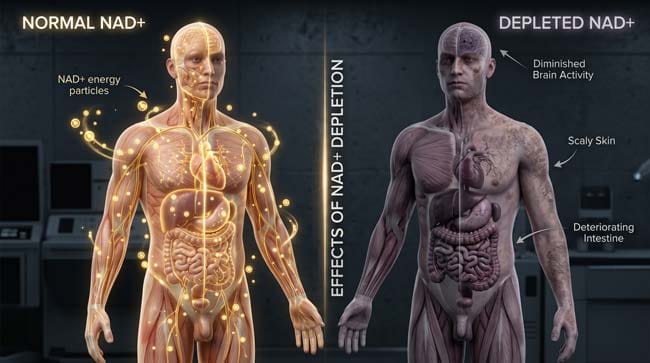
My 3D Discovery: Rendering a Body Running Out of Power
When I was building the NAD+ depletion model for this simulation, the most compelling visualization was comparing a fully powered cell versus a NAD+-depleted cell side by side. In a healthy cell, the mitochondria pulse with a constant golden light — ATP being produced in a steady, rhythmic cycle. In a Niacin-deficient cell, that pulse slows, stutters, and eventually flatlines.
What makes Vitamin B3 deficiency uniquely dramatic in 3D is that the failure is not localized. It spreads across every tissue simultaneously — because every cell in your body depends on NAD+ for energy.
3D Observation: The most visually striking sequence in this simulation is watching the deficiency propagate from the cellular level outward. It starts as a flickering in individual mitochondria. Then entire cells dim. Then tissue-level function deteriorates. Then organ systems begin to fail — skin, intestine, brain, in succession. It looks less like a disease and more like a building losing power floor by floor, section by section, until the entire structure goes dark.
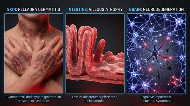
Stage 1: How Vitamin B3 Deficiency Develops — The Cellular Mechanism
Understanding why Vitamin B3 deficiency is so damaging requires understanding what NAD+ actually does inside your cells.
NAD+ (Nicotinamide Adenine Dinucleotide) is not a fuel source itself — it is an electron carrier. During cellular respiration, NAD+ accepts electrons from glucose breakdown products, becoming NADH. NADH then donates these electrons to the mitochondrial electron transport chain, which uses the energy to produce ATP.
Without sufficient Niacin to maintain NAD+ levels, this entire chain breaks down.
Secondary roles of NAD+ that also fail:
- DNA repair — NAD+ is consumed by PARP enzymes that repair DNA strand breaks. Without it, DNA damage accumulates uncorrected.
- Gene expression regulation — Sirtuins (longevity proteins) require NAD+ to function. Their activity collapses with deficiency.
- Calcium signaling — NAD+ derivatives regulate intracellular calcium — critical for muscle contraction and nerve function.
| NAD+ Function | Normal State | Deficiency State | Consequence |
|---|---|---|---|
| ATP Production | Continuous mitochondrial cycling | Slowing then halting | Cellular energy failure |
| DNA Repair | Active PARP enzyme function | PARP activity collapses | DNA damage accumulation |
| Sirtuin Activity | Gene regulation maintained | Sirtuins inactive | Accelerated cellular aging |
| Calcium Signaling | Regulated ion flow | Dysregulated | Nerve and muscle dysfunction |
Who develops Vitamin B3 deficiency:
- People consuming corn-based diets without nixtamalization
- Individuals with alcohol use disorder — alcohol interferes with Niacin absorption and metabolism
- People with Hartnup disease — a genetic disorder affecting Tryptophan absorption (Tryptophan is converted to Niacin in the body)
- People with carcinoid syndrome — tumors divert Tryptophan away from Niacin synthesis
- Severe malnutrition cases generally
According to the National Institutes of Health (NIH), the recommended daily intake of Niacin for adults is 14–16 mg per day — an amount easily obtained from a varied diet including meat, fish, nuts, and legumes, but critically absent in unprocessed corn-dominant diets. NIH: Niacin Fact Sheet
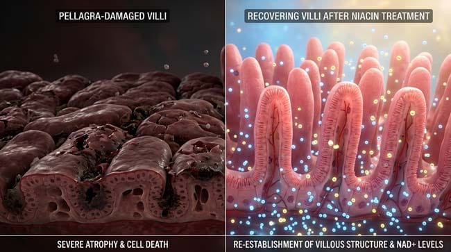
Stage 2: The Three Systems That Fail First — Skin, Gut, Brain
Vitamin B3 deficiency does not affect all tissues equally. The tissues that fail first are those with the highest cell turnover rates — because rapidly dividing cells require the most NAD+ for energy and DNA repair.
System 1: Skin — Dermatitis
Skin cells divide continuously. In our 3D simulation, the skin surface shader begins changing at approximately 30% NAD+ depletion — the smooth, translucent surface texture giving way to a rough, thickened appearance.
The characteristic Pellagra skin rash has three distinctive features that I rendered in detail:
Photosensitivity — Sun-exposed areas are affected first and most severely. In the simulation, UV radiation hitting NAD+-depleted skin cells causes DNA damage that cannot be repaired — the cells die and trigger inflammatory responses that produce the rash pattern.
Symmetry — The rash appears symmetrically — both hands, both forearms, the back of the neck. In the 3D model, this bilateral pattern reflects the equal UV exposure of symmetrical body surfaces.
Casal’s Necklace — The distinctive rash around the neck, named after the Spanish physician Gaspar Casal who first described Pellagra in 1735. In the 3D render, this appears as a dark, clearly demarcated band encircling the lower neck — one of the most visually recognizable signs of advanced Niacin deficiency.
System 2: Digestive Tract — Diarrhea
The intestinal lining is one of the fastest-renewing tissues in the body — completely replaced every 3–5 days. This makes it extremely sensitive to NAD+ depletion.
In our 3D simulation, intestinal villi — the tiny finger-like projections that massively increase surface area for nutrient absorption — begin shrinking and flattening at moderate NAD+ depletion levels.
The consequences compound rapidly:
- Flattened villi → reduced absorption surface area
- Reduced absorption → worse nutritional status overall
- Worse nutritional status → accelerated NAD+ depletion
- Accelerated depletion → faster villi deterioration
This self-reinforcing cycle is why Pellagra patients deteriorate so rapidly once digestive symptoms begin — the digestive failure itself prevents adequate nutrient absorption even if the diet improves.
System 3: Brain and Nervous System — Dementia
Neurons are among the most NAD+-dependent cells in the body. They cannot divide to replace themselves (unlike skin or intestinal cells), making neurological damage the most potentially permanent consequence of severe deficiency.
In our 3D neurological model, NAD+ depletion produces four visible changes:
Neurotransmitter synthesis failure — Serotonin, dopamine, and other neurotransmitters require NAD+-dependent enzymatic reactions. As production drops, mood disorders and cognitive impairment develop before structural damage is visible.
Myelin deterioration — The fatty myelin sheath surrounding nerve fibers begins thinning in the simulation. This slows nerve conduction velocity — shown in the animation as signal pulses traveling progressively more slowly along nerve pathways.
Synaptic weakening — NAD+-dependent maintenance of synaptic connections fails. In the viewport, active synaptic connections shown as bright nodes begin dimming and eventually going dark — representing the literal loss of neural connectivity.
Mitochondrial failure in neurons — Neurons have extremely high mitochondrial density due to their energy demands. In the 3D model, mitochondrial failure in neurons is particularly dramatic — entire sections of neural tissue losing their energy supply simultaneously.
| Neurological Symptom | Early Stage | Advanced Stage |
|---|---|---|
| Mood changes | Anxiety, irritability | Severe depression |
| Cognitive function | Mild confusion, memory issues | Dementia, disorientation |
| Motor control | Mild coordination issues | Significant impairment |
| Consciousness | Generally maintained | Delirium in severe cases |
According to the Mayo Clinic, neurological symptoms of Pellagra can sometimes be mistaken for psychiatric disorders — leading to misdiagnosis in populations where the dietary cause is not immediately considered. Mayo Clinic: Pellagra Overview
Stage 3: Treatment and Recovery — What 3D Shows Us About Healing
One of the most remarkable aspects of Vitamin B3 deficiency is how rapidly the body can recover with proper treatment — at least in the early stages.
Treatment Protocol: The standard treatment is Niacin supplementation — typically 100–300mg per day in divided doses for several weeks, followed by ongoing dietary correction.
In our 3D recovery simulation, the healing sequence is almost the reverse of the damage sequence:
Day 1–3: NAD+ levels begin rising. Mitochondrial function improves. Cellular energy production restarts. In the viewport, the mitochondrial pulse returns — dim at first, then strengthening.
Week 1–2: Skin inflammation begins resolving. The rough, darkened surface texture in the shader begins softening. New healthy skin cells, now with adequate NAD+ for DNA repair, begin replacing damaged tissue.
Week 2–3: Intestinal villi begin recovering height and density. Nutrient absorption improves. The self-reinforcing cycle of digestive failure begins reversing.
Week 3–8: Neurological symptoms begin improving — mood, cognitive clarity, coordination. The recovery here is slower and less complete than skin and digestive recovery.
| Body System | Response to Treatment | Recovery Timeline |
|---|---|---|
| Skin (Dermatitis) | Rapid — inflammation resolves quickly | 1–3 weeks |
| Digestive (Diarrhea) | Moderate — villi regrowth takes time | 2–4 weeks |
| Brain (Dementia) | Slow — neurological repair is complex | Weeks to months |
| Severe neurological damage | Partial — may be permanent | Incomplete recovery |
FAQ: Vitamin B3 Deficiency Symptoms
Q1: How do I know if I have Vitamin B3 deficiency? Early symptoms are nonspecific — fatigue, mild digestive discomfort, and skin sensitivity to sunlight. The appearance of the characteristic symmetrical rash on sun-exposed areas, combined with digestive and cognitive symptoms, is the classic clinical indicator. A blood test measuring Niacin metabolites can confirm deficiency. Always consult a qualified healthcare provider for diagnosis.
Q2: Can you get too much Vitamin B3? Yes. High-dose Niacin supplementation (above 35mg per day from supplements) can cause Niacin flush — a temporary redness, warmth, and tingling of the skin caused by vasodilation. At very high doses (1,000mg+ used therapeutically for cholesterol management), liver toxicity is a risk. The tolerable upper intake level set by the NIH is 35mg per day from supplements for adults.
Q3: What foods are highest in Vitamin B3? The richest dietary sources include chicken breast (~11mg per 100g), tuna (~22mg per 100g), beef liver (~17mg per 100g), peanuts (~14mg per 100g), and brown rice (~5mg per 100g). Tryptophan in protein-rich foods also contributes — the body converts approximately 60mg of dietary Tryptophan into 1mg of Niacin.
Q4: Is Pellagra still common today? In developed countries with fortified food supplies, clinical Pellagra is rare. In regions where unprocessed corn is a dietary staple without supplementation — parts of sub-Saharan Africa, rural India, and some refugee populations — Pellagra remains a genuine public health concern. The WHO lists it as a priority micronutrient deficiency condition.
Q5: Does alcohol cause Vitamin B3 deficiency? Chronic alcohol use is one of the leading causes of Pellagra in developed countries. Alcohol interferes with Niacin absorption in the intestine, disrupts the liver’s conversion of Tryptophan to Niacin, and often displaces nutritious food in the diet. People with severe alcohol use disorder frequently present with multiple B-vitamin deficiencies simultaneously.
Conclusion: The Four Ds — A Warning Written Across Every Body System
Vitamin B3 deficiency is medicine’s most visually complete nutritional disorder — because it does not hide. It writes its symptoms across the skin in darkened patches, announces itself through digestive collapse, and ultimately reaches the brain itself.
In 3D, rendering the progression from cellular NAD+ depletion to full Pellagra — and then watching the recovery when Niacin is restored — is one of the most complete demonstrations of how a single molecule can determine the functional integrity of the entire human body.
The good news is that prevention requires only dietary variety, and treatment works quickly. The damage of centuries of Pellagra epidemics came not from lack of a cure, but from lack of understanding of the cause — a lesson that modern nutritional science has permanently resolved.
Further Study & External Research
3D Simulation Specs & Observations
| 3D Component | Technical Visual Setting | Observation from Viewport |
|---|---|---|
| Framerate | 120 FPS High-Speed | Captured NAD+ depletion and tissue deterioration progression |
| Material/Shader | Subsurface Scattering (SSS) | Simulating skin texture changes from healthy to Pellagra-affected |
| Physics Engine | Volumetric Particle System | Visualized NAD+ molecules as glowing energy particles |
| Goal | Educational / Science Visualization | Research-referenced 3D breakdown of Vitamin B3 deficiency |
Read more on What Happens If You Only Eat Corn?
Pingback: What Happens If You Only Eat Corn? 3D Body Breakdown