
The Creator’s Note & Disclaimer: As a 3D artist at WhatIfBody3D, I rendered this scenario at 120 FPS. Our models explore why you should never pop a pimple in the triangle of death — visualizing the facial danger triangle’s vascular anatomy, bacterial translocation pathways, cavernous sinus thrombosis mechanics, and the life-threatening cascade that can follow infected pimple manipulation in this specific facial zone. This visualization is part of our “What If” series and is for educational and informational purposes only, as stated in our About Page.
Quick Answer: Why Should You Never Pop a Pimple in the Triangle of Death? (The Atomic Answer)
Why you should never pop a pimple in the triangle of death comes down to one extraordinary anatomical fact — the veins draining this area of your face have a direct connection to the inside of your skull.
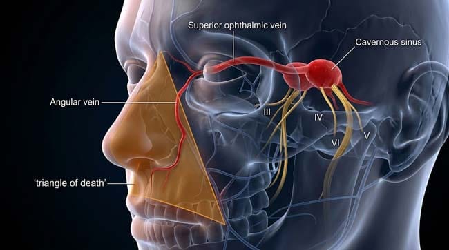
- The Triangle: The “Triangle of Death” (also called the Danger Triangle) is the facial region bounded by the corners of the mouth and the bridge of the nose — covering the nasal area, upper lip, and nasolabial folds.
- The Vascular Connection: Veins in this region drain backward into the cavernous sinus — a large venous space inside the skull, directly adjacent to critical cranial nerves and the brain.
- The Risk: Unlike most facial veins, the veins in the danger triangle lack valves — meaning blood (and anything in it, including bacteria) can flow in either direction — forward toward the face or backward toward the brain.
- The Cascade: Squeezing an infected pimple in this zone can force bacteria directly into the valveless venous system — potentially causing cavernous sinus thrombosis (CST) — an infected blood clot in the skull that carries a 30% mortality rate even with modern treatment.
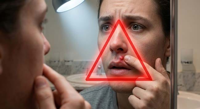
My 3D Discovery: Rendering the “Highway to the Brain”
When I was building the facial vascular model for this simulation, the most striking realization was just how direct the anatomical connection is between the danger triangle’s superficial veins and the intracranial venous system. In most areas of the face, venous drainage flows downward and outward — away from the brain. In the danger triangle, the venous anatomy is fundamentally different.
In the 3D viewport, I rendered the facial venous system as a branching network of tubes — showing how the angular vein, superior ophthalmic vein, and inferior ophthalmic vein form a direct pathway from the corner of the nose to the cavernous sinus inside the skull. Without valves to enforce directional flow, pressure from squeezing a pimple shown forcing flow backward — directly up this anatomical highway toward the brain.
3D Observation: The most visually alarming sequence in this simulation is the bacterial translocation animation. A single squeeze of an infected pimple shown creating a pressure wave in the surrounding tissue — forcing infected material into the adjacent venous network. Bacteria rendered as red particles shown entering the vein and immediately flowing in two directions simultaneously — forward and backward. The backward flow shown traveling up the angular vein, through the ophthalmic veins, and into the cavernous sinus within seconds. The cavernous sinus shown as a large pool-like space suddenly receiving a bacterial inoculation — the infection beginning there, deep inside the skull, before any external symptom appears.
Stage 1: The Danger Triangle — Anatomy of a Vascular Hazard
What and Where is the Triangle of Death?
The facial danger triangle is bounded by three anatomical landmarks:
- Left corner of the mouth
- Right corner of the mouth
- Bridge of the nose (nasion)
This triangular region covers:
- The upper lip
- Both nasolabial folds
- The nose — particularly the nasal tip and alae
- The philtrum (groove between nose and upper lip)
- The perinasal area
In our 3D facial model, I rendered this triangle as an overlay on the face — highlighting the skin, subcutaneous tissue, and underlying vascular structures within this zone.
Why This Specific Area?
The danger triangle’s hazard is purely anatomical — defined entirely by the vascular drainage pattern of this facial region. The key structure is the angular vein — a superficial vein running along the side of the nose from the inner corner of the eye downward.
The Angular Vein’s Dangerous Connections:
In our 3D vascular model, I traced the complete connection:
Step 1: The angular vein runs along the lateral nose — receiving tributaries from the nasal tip, upper lip, and nasolabial fold
Step 2: At the medial canthus (inner eye corner), the angular vein connects to the superior ophthalmic vein — shown as a larger vessel running backward through the orbit
Step 3: The superior ophthalmic vein passes through the superior orbital fissure — a gap in the skull bones at the back of the eye socket
Step 4: It drains directly into the cavernous sinus — a large dural venous sinus sitting at the base of the skull, immediately adjacent to the pituitary gland, cranial nerves III, IV, V, and VI, and the internal carotid artery
The Absence of Valves:
Most peripheral veins contain one-way valves — flap-like structures that ensure blood flows in one direction only (toward the heart). The ophthalmic veins and their connections in the danger triangle are notably avalvular — they lack these directional valves.
In our 3D valve model, I showed a comparison:
- Normal peripheral vein: shown with valve leaflets opening as blood flows toward heart and closing to prevent backflow
- Danger triangle vein: shown as a smooth tube with no valve structures — blood shown flowing freely in either direction depending on pressure gradients
This avalvular anatomy is the fundamental reason the danger triangle is dangerous — it means pressure from squeezing, infection, or thrombosis can drive flow in any direction, including directly toward the cavernous sinus.
| Anatomical Structure | Location | Normal Function | Danger Triangle Role |
|---|---|---|---|
| Angular vein | Along lateral nose | Facial venous drainage | Primary bacterial entry point |
| Superior ophthalmic vein | Through orbit | Orbital venous drainage | Direct conduit to cavernous sinus |
| Inferior ophthalmic vein | Lower orbit | Orbital drainage | Secondary conduit |
| Superior orbital fissure | Back of eye socket | Venous passage through skull | Gateway to intracranial space |
| Cavernous sinus | Base of skull | Dural venous drainage | Target of infection — adjacent to brain |
According to the American Journal of Neuroradiology, the cavernous sinus is one of the most strategically dangerous locations for infection in the entire body — its proximity to cranial nerves III, IV, V1, V2, and VI means that thrombosis or infection in this space produces a characteristic constellation of cranial nerve deficits that is clinically unmistakable. AJNR: Cavernous Sinus Anatomy and Pathology
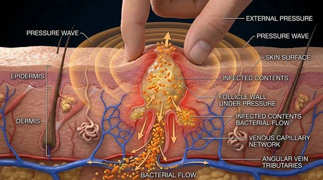
Stage 2: The Infected Pimple — What Happens When You Squeeze
Normal Pimple Biology:
A pimple (acne vulgaris) in the danger triangle is essentially a follicular abscess — a collection of bacteria, dead white blood cells, sebum, and keratin debris within a hair follicle unit.
In our 3D follicular model, I rendered a typical danger triangle pimple:
- Follicle wall: Shown as a cylindrical structure containing the infection
- Contents: Staphylococcus aureus and Cutibacterium acnes shown as the primary bacterial species, surrounded by neutrophils, sebum, and keratin
- Surrounding tissue: Inflamed, with dilated capillaries and attracted immune cells
The Squeeze Mechanics:
When pressure is applied to squeeze the pimple, our 3D pressure simulation showed three simultaneous events:
Event 1 — Follicle Wall Rupture The follicle wall shown failing under applied pressure — the weakest point (usually the isthmus between the follicle and the dermis) shown tearing. Infected contents shown being forced outward — some toward the skin surface, but critically, some driven deeper into the dermis.
Event 2 — Dermal Inoculation Bacteria forced deeper into the dermis shown entering the rich capillary network of the upper dermis. From here, bacteria shown accessing the venous drainage system — entering the angular vein tributaries.
Event 3 — Retrograde Venous Flow The pressure wave from squeezing shown creating a momentary pressure spike in the venous system. In the avalvular danger triangle veins, this pressure spike shown driving a pulse of bacterial-laden blood backward — up the angular vein, through the ophthalmic veins, toward the cavernous sinus.
The Bacterial Species:
In our molecular pathogen model, I showed the two primary species involved in danger triangle pimple infections:
Staphylococcus aureus (shown as orange clusters)
- Most common cause of cavernous sinus thrombosis following facial infection
- Produces coagulase — shown as an enzyme that triggers local clot formation
- Produces toxins that damage vascular endothelium — shown as molecules disrupting vessel wall integrity
Cutibacterium acnes (shown as small blue rods)
- Less virulent but more prevalent in typical acne
- Can trigger inflammatory cascades that promote thrombosis even without direct bacterial invasion
| Squeeze Event | 3D Visual | Clinical Consequence |
|---|---|---|
| Follicle wall rupture | Container wall shown tearing under pressure | Infected material dispersed into dermis |
| Dermal inoculation | Bacteria shown entering capillary network | Bacteremia risk — bacteria in bloodstream |
| Retrograde venous flow | Pressure wave shown driving flow backward | Bacteria potentially reaching cavernous sinus |
| Vascular endothelial damage | Staphylococcal toxins shown disrupting vessel wall | Promotes thrombus formation |
| Local immune response | Neutrophil flood shown at rupture site | Inflammation amplifies pressure, worsening retrograde flow |
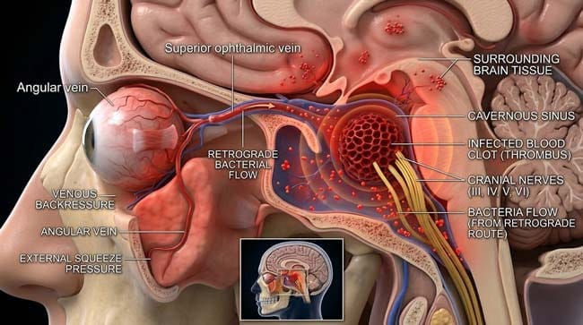
Stage 3: Cavernous Sinus Thrombosis — The Life-Threatening Cascade
What is Cavernous Sinus Thrombosis (CST)?
CST is an infected blood clot within the cavernous sinus — one of the most serious infectious complications in medicine. In our 3D cavernous sinus model, I rendered the sinus as a large trabeculated (honeycomb-structured) venous space at the base of the skull.
The Structures Adjacent to the Cavernous Sinus:
In the 3D model, I showed the extraordinary density of critical structures immediately surrounding the cavernous sinus:
- Cranial Nerve III (Oculomotor) — controls eye movement
- Cranial Nerve IV (Trochlear) — controls superior oblique eye muscle
- Cranial Nerve V1 (Ophthalmic branch of trigeminal) — sensation to forehead and eye
- Cranial Nerve V2 (Maxillary branch of trigeminal) — sensation to cheek and upper lip
- Cranial Nerve VI (Abducens) — controls lateral eye movement
- Internal Carotid Artery — primary blood supply to the brain
The CST Progression:
Phase 1 — Bacterial Arrival (Hours 0–24) Bacteria shown arriving at the cavernous sinus via retrograde venous flow. Initial infection shown as small bacterial colonies establishing in the sinus trabeculations — producing local inflammation.
Phase 2 — Thrombosis Formation (Hours 24–72) Bacterial coagulase and endothelial damage shown triggering clot formation within the sinus. The thrombus shown expanding progressively — blocking venous outflow from the orbit and brain.
Phase 3 — Cranial Nerve Involvement (Days 2–5) As inflammation and clot expand, shown compressing adjacent cranial nerves. The characteristic CST symptom triad begins:
- Proptosis (eye bulging) — shown as orbital venous congestion pushing the eye forward
- Chemosis (conjunctival swelling) — shown as orbital congestion affecting the conjunctiva
- Ophthalmoplegia (loss of eye movement) — shown as cranial nerve III, IV, and VI compression
Phase 4 — Intracranial Extension (Days 3–7 without treatment) Without aggressive treatment, shown thrombus extending to adjacent sinuses — the superior sagittal sinus and transverse sinuses. Meningeal involvement shown producing bacterial meningitis. Cerebral venous infarction shown as brain tissue damage from venous outflow obstruction.
The Statistics:
- Pre-antibiotic era mortality: >80%
- Current mortality with aggressive treatment: approximately 30%
- Permanent neurological deficit in survivors: approximately 50%
These numbers explain why “Triangle of Death” is not hyperbole — it is a reflection of the genuine anatomical risk.
| CST Stage | Timeline | 3D Visualization | Clinical Signs |
|---|---|---|---|
| Bacterial arrival | Hours 0–24 | Bacteria shown in cavernous sinus | None yet — pre-symptomatic |
| Thrombosis | Hours 24–72 | Clot forming in sinus space | Facial pain, fever beginning |
| Cranial nerve compression | Days 2–5 | Nerves shown compressed by expanding thrombus | Proptosis, chemosis, ophthalmoplegia |
| Intracranial extension | Days 3–7 | Thrombus shown extending to adjacent sinuses | Meningism, altered consciousness |
| Without treatment | Days 5–14 | Multi-sinus involvement, cerebral infarction | Coma, death |
According to the Journal of Neurology, Neurosurgery and Psychiatry, cavernous sinus thrombosis secondary to facial infection — including acne — remains a medical emergency associated with significant mortality and morbidity despite modern antibiotic therapy and anticoagulation treatment. JNNP: Cavernous Sinus Thrombosis — Clinical Review
FAQ: Why You Should Never Pop a Pimple in the Triangle of Death
Q1: How common is cavernous sinus thrombosis from pimple popping? Cavernous sinus thrombosis is rare — with an estimated incidence of approximately 1 per 500,000 people annually. However, it is significantly more common than most rare conditions because its primary triggers (facial infections, dental infections, sinusitis) are extremely common. The risk from a single pimple-popping event is low but non-zero — and given the severity of outcomes when it does occur, avoidance is strongly recommended regardless of the absolute risk percentage.
Q2: Is it ever safe to pop a pimple in the danger triangle? Dermatologists recommend against squeezing any inflamed pimple anywhere on the face — particularly in the danger triangle. If a pimple in this region becomes severely painful, rapidly enlarging, or shows signs of deep infection (warmth, redness spreading beyond the pimple border, fever), medical evaluation is appropriate. Professional extraction under sterile conditions by a dermatologist carries significantly lower infection spread risk than home squeezing.
Q3: What are the warning signs of cavernous sinus thrombosis after facial manipulation? Warning signs requiring immediate emergency care: severe headache that is different from any previous headache, fever, one or both eyes protruding abnormally, reduced or double vision, drooping eyelid, swelling around the eye, stiff neck, or altered consciousness occurring days after squeezing a pimple in the danger triangle area. These symptoms within 1–2 weeks of facial infection or manipulation warrant emergency evaluation.
Q4: Does the same risk apply to nose picking in the danger triangle area? Yes — nose picking traumatizes the nasal vestibule mucosa, which is also within the danger triangle’s vascular drainage zone. This is why nasal vestibulitis (infection from nose picking) can, in rare cases, progress to cavernous sinus thrombosis through the same venous pathway. The risk is higher when the nasal mucosa is broken, infected, and repeatedly traumatized.
Q5: Are there safe ways to treat pimples in the danger triangle? Yes — topical treatments that do not involve mechanical manipulation are safe. Benzoyl peroxide, salicylic acid, and topical retinoids address the bacterial and comedonal components without creating pressure that drives bacteria into the venous system. For persistent or inflamed lesions, a dermatologist can perform safe extractions or prescribe oral antibiotics that treat the infection systemically without the mechanical risk of home squeezing.
Conclusion: The Most Dangerous Square Inch on Your Face
The triangle of death is not a myth, not an exaggeration, and not a medical urban legend. It is a precise anatomical description of a facial region where the vascular drainage pattern creates a direct, valveless connection between skin surface infections and the inside of the skull.
In 3D, rendering the complete pathway — from a squeezed pimple’s pressure wave forcing bacteria into the angular vein, through the avalvular ophthalmic veins, into the cavernous sinus, and watching the subsequent thrombosis compress the cranial nerves packed into that small intracranial space — makes the anatomy immediately and viscerally clear.
Most pimple-popping events in the danger triangle produce nothing worse than a slightly worse pimple. The anatomy does not care about statistics. The pathway exists every time. The valves are always absent. The cavernous sinus is always adjacent.
Think twice before you squeeze.
Further Study & External Research
3D Simulation Specs & Observations
| 3D Component | Technical Visual Setting | Observation from Viewport |
|---|---|---|
| Framerate | 120 FPS High-Speed | Captured bacterial translocation dynamics and venous flow mechanics |
| Material/Shader | Subsurface Scattering (SSS) | Simulating facial skin tissue, vessel wall translucency, and sinus space |
| Physics Engine | Fluid Dynamics + Volumetric Particle System | Visualized retrograde venous flow, bacterial particle movement, thrombus formation |
| Goal | Educational / Science Visualization | Research-referenced 3D breakdown of facial danger triangle vascular anatomy |
Read more on What Happens When You Pop a Pimple?
Pingback: What Happens When You Pop a Pimple? The 3D Skin Science