
The Creator’s Note & Disclaimer: As a 3D artist at WhatIfBody3D, I rendered this scenario at 120 FPS. Our models explore how your body fights airborne viruses — visualizing the innate immune response, interferon signaling cascade, natural killer cell deployment, T cell activation, and antibody production against inhaled respiratory pathogens. This visualization is part of our “What If” series and is for educational and informational purposes only, as stated in our About Page.
Quick Answer: How Does Your Body Fight Airborne Viruses? (The Atomic Answer)
How does your body fight airborne viruses? Through a two-wave immune response — a rapid, non-specific innate response that activates within hours, followed by a precise, targeted adaptive response that takes days to mobilize but eliminates the virus with extraordinary specificity.
- Wave 1 — Innate Immunity (Hours 0–72): The moment infected cells detect viral RNA, they broadcast emergency signals — interferons — that warn surrounding cells and recruit Natural Killer cells, macrophages, and neutrophils to the infection site.
- Wave 2 — Adaptive Immunity (Days 3–14): T lymphocytes and B lymphocytes are activated, producing virus-specific cytotoxic T cells that destroy infected cells and plasma B cells that manufacture millions of targeted antibodies per second.
- The Memory: After the infection is cleared, memory T cells and memory B cells persist for years — providing rapid protection against future exposure to the same virus.
- The Timeline: A typical respiratory viral infection is detected by the innate system within 4–12 hours, controlled by the innate response within 3–5 days, and eliminated by the adaptive response within 7–14 days in a healthy individual.
My 3D Discovery: Rendering the “Two-Army Defense”
When I was building the immune response model for this simulation, the most visually striking element was the contrast between the two immune waves. The innate response — rendered in warm amber and red tones — looks like a rapid, somewhat chaotic emergency response: macrophages rushing to the site, interferons spreading in all directions, natural killer cells destroying anything that looks infected.
The adaptive response — rendered in cool blue and silver tones — looks completely different: precise, deliberate, targeted. T cells shown moving with purpose toward specific infected cells. B cells shown in lymph nodes producing identical antibodies in perfect assembly-line fashion. The contrast between the two systems in the same simulation makes the division of labor visually obvious.
3D Observation: The most scientifically remarkable sequence in this simulation is the T cell activation moment. A naive T cell — shown as a small, unremarkable sphere — encounters a dendritic cell presenting a viral peptide on an MHC molecule. The moment of recognition is shown as a precise molecular lock-and-click — the T cell receptor finding its exact complementary peptide-MHC complex. In the animation, this triggers an immediate transformation: the T cell begins dividing rapidly, shown as a single cell expanding into a clone of thousands of identical cells — all carrying the same receptor, all targeted at the same viral protein. It is one of the most elegant examples of biological precision in the entire immune system.
Stage 1: Innate Immunity — The First Responders
The innate immune response is the body’s immediate, non-specific defense against viral infection. It activates within hours of viral detection and does not require prior exposure to the specific virus — it responds to general viral signatures that all viruses share.
The Interferon Response — The Body’s Viral Alarm System
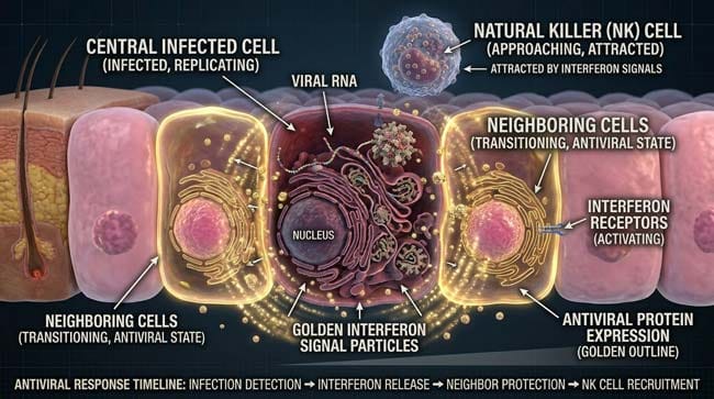
The first and most critical component of the innate antiviral response is the interferon (IFN) signaling cascade. When an infected respiratory epithelial cell detects viral RNA through its Pattern Recognition Receptors (PRRs) — specifically RIG-I and MDA5 sensors — it immediately begins producing and secreting Type I interferons (IFN-α and IFN-β).
In our 3D signaling model, I rendered interferons as bright golden signal particles — shown radiating outward from the infected cell in all directions, reaching neighboring cells within minutes.
What Interferons Do:
To surrounding uninfected cells: Interferon binding triggers an antiviral state — shown in the animation as neighboring cells changing from their normal pink color to a golden-outlined protected state. This involves upregulating hundreds of Interferon-Stimulated Genes (ISGs) that:
- Degrade viral RNA before it can be translated
- Shut down protein synthesis to prevent viral replication
- Increase MHC Class I presentation to alert cytotoxic T cells
- Activate apoptosis pathways — shown as cells preparing to self-destruct if infected
To immune cells: Interferons shown as attracting and activating Natural Killer (NK) cells and macrophages — drawing them toward the infection site through chemokine gradients.
Interferon Evasion by Viruses: In a parallel simulation track, I showed why some respiratory viruses — particularly influenza and SARS-CoV-2 — cause more severe illness than others. Both viruses produce proteins that specifically block interferon production:
- Influenza NS1 protein — shown as a molecular blocker that binds and inactivates RIG-I
- SARS-CoV-2 ORF3b protein — shown as blocking the signaling cascade downstream of interferon production
In the animation, this interferon evasion appears as the golden alarm signal being suppressed — the infected cell producing little or no interferon, leaving surrounding cells without warning and buying the virus additional replication time.
| Interferon Type | Producer Cell | 3D Color | Primary Effect | Timing |
|---|---|---|---|---|
| IFN-α | Plasmacytoid dendritic cells | Bright gold | Systemic antiviral state | 4–8 hours |
| IFN-β | Infected epithelial cells | Amber | Local antiviral state | 4–6 hours |
| IFN-γ | NK cells and T cells | Orange | Macrophage activation | 12–24 hours |
| IFN-λ | Respiratory epithelial cells | Yellow-gold | Mucosal antiviral defense | 6–12 hours |
Natural Killer Cells — The Early Cytotoxic Response
Natural Killer (NK) cells are innate immune cells that destroy virally infected cells without requiring prior sensitization. In our 3D model, NK cells shown as medium-sized spherical cells moving rapidly through tissue — scanning surrounding cells for signs of infection.
How NK Cells Identify Infected Cells:
Normal healthy cells display MHC Class I molecules on their surface — shown as small flag-like proteins. NK cells have inhibitory receptors that bind to MHC Class I — this binding signal tells the NK cell “this is a healthy cell, do not kill.”
Many viruses — to evade cytotoxic T cells — downregulate MHC Class I expression. In the 3D model, infected cells shown with reduced or absent MHC flags. When an NK cell encounters a cell with missing MHC flags, the inhibitory signal is absent — the NK cell receives no “don’t kill” signal and activates its killing mechanism.
The NK Cell Kill Sequence:
- NK cell shown making contact with infected cell
- Cytotoxic granules — shown as glowing red vesicles — released toward the target cell
- Perforin molecules shown punching holes in the target cell membrane
- Granzymes shown entering through the perforin pores and triggering apoptosis
- Target cell shown beginning to collapse and fragment — the infection destroyed before viral replication completes
According to the National Institute of Allergy and Infectious Diseases (NIAID), the interferon response and NK cell activity represent the most critical determinants of early viral infection outcome — individuals with impaired interferon production or NK cell function experience dramatically more severe respiratory viral illness. NIAID: Innate Immunity and Viral Infection
Stage 2: The Bridge — Dendritic Cells Linking Innate to Adaptive
The transition from the innate to the adaptive immune response depends on a critical cell type — the dendritic cell — which acts as the intelligence link between the two systems.
How Dendritic Cells Work:
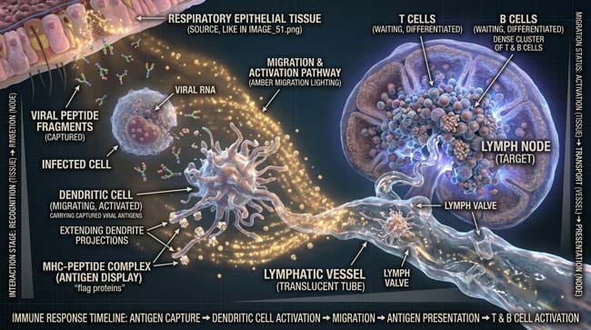
Dendritic cells are professional antigen-presenting cells — they patrol tissues, engulf infected cells and viral particles, process them into peptide fragments, and travel to the nearest lymph node to present these fragments to T lymphocytes.
In our 3D dendritic cell model, this process appears as:
Step 1 — Sampling Dendritic cells shown extending long projections (dendrites) across the respiratory epithelium — sampling the environment for signs of infection. In the simulation, these dendrites shown capturing viral particles and infected cell debris.
Step 2 — Processing Inside the dendritic cell, viral proteins shown being broken down into peptide fragments — shown as the complex viral protein being cut into small pieces by proteases in the endosome.
Step 3 — MHC Loading Peptide fragments loaded onto MHC Class II molecules — shown as the peptide piece fitting into the groove of the MHC molecule on the dendritic cell surface.
Step 4 — Migration The dendritic cell shown migrating from the respiratory tissue through lymphatic vessels to the nearest lymph node — carrying viral peptide-MHC complexes as evidence of the infection.
Step 5 — T Cell Presentation In the lymph node, the dendritic cell shown scanning through thousands of naive T cells — displaying its viral peptide-MHC complex until it finds the T cell with the complementary receptor.
The Lymph Node — Adaptive Response Headquarters:
In our 3D lymph node model, I showed the node as a dense cluster of immune cells — naive T cells and B cells constantly circulating through the node, each with unique receptors that recognize different molecular targets.
The dendritic cell’s arrival with viral antigen shown as triggering a cascade of recognition events — multiple T cells shown finding their complementary peptide-MHC matches simultaneously, initiating clonal expansion across multiple T cell specificities.
Stage 3: Adaptive Immunity — The Precision Strike Force
The adaptive immune response takes longer to mobilize — 3–7 days for initial activation, 7–14 days for full deployment — but it is overwhelmingly more powerful and specific than the innate response.
Cytotoxic T Cells — Targeted Cell Destroyers
CD8+ cytotoxic T lymphocytes (CTLs) are the adaptive immune system’s primary antiviral effector cells. Once activated by dendritic cell presentation, they undergo clonal expansion — dividing rapidly to produce thousands of identical copies, all carrying the same virus-specific T cell receptor.
In our 3D CTL model, activated T cells shown in the lymph node as a single cell dividing into two, then four, then eight — the exponential expansion shown accelerating over days until the lymph node contains tens of thousands of identical T cells, all specific for the same viral peptide.
How CTLs Destroy Infected Cells:
Once released from the lymph node, CTLs travel through the bloodstream to the infection site — shown in the simulation as blue-tinted cells streaming through blood vessels toward the respiratory tract.
At the infection site, CTLs shown scanning epithelial cells — examining their MHC Class I displays. Infected cells display viral peptide fragments on MHC Class I — shown as a different flag configuration that the CTL receptor recognizes as foreign.
The Kill Sequence:
- CTL shown making contact with infected cell
- T cell receptor binding to viral peptide-MHC complex — shown as precise molecular lock-and-click
- CTL activating and releasing cytotoxic granules — perforin and granzyme
- Infected cell shown undergoing controlled apoptosis — fragmenting cleanly without releasing inflammatory contents
- CTL detaching and moving to next infected cell — shown as a systematic, methodical clearing operation
B Cells and Antibodies — The Molecular Neutralization System
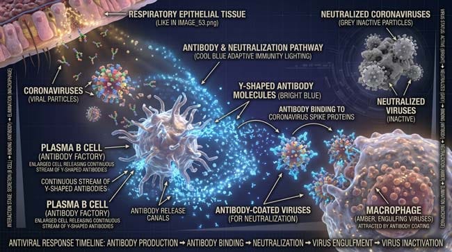
While CTLs destroy infected cells, B lymphocytes produce antibodies — Y-shaped protein molecules that neutralize free viral particles before they can infect new cells.
In our 3D B cell model, virus-specific B cells shown in the lymph node being activated by viral antigen and T helper cell signals simultaneously. Activated B cells transform into plasma cells — shown as enlarged, factory-like cells with dramatically expanded endoplasmic reticulum — optimized for antibody production.
A single plasma cell can produce 2,000 antibody molecules per second — shown in the animation as a continuous stream of Y-shaped proteins being released from the plasma cell into the surrounding tissue.
How Antibodies Neutralize Viruses:
In our 3D molecular model, antibodies shown binding to the viral spike protein — the same protein that binds to ACE2 receptors. Antibody binding shown physically blocking the receptor-binding domain — the virus can no longer attach to cells.
Additional antibody mechanisms shown:
- Neutralization — antibodies coating virus shown preventing ACE2 binding
- Opsonization — antibody-coated viruses shown being recognized and engulfed by macrophages
- Complement activation — antibodies shown triggering the complement cascade that destroys viral particles
| Adaptive Immune Component | Activation Time | Primary Function | 3D Visualization |
|---|---|---|---|
| CD8+ Cytotoxic T cells | Days 3–5 | Destroy infected cells | Blue cells scanning and killing infected epithelium |
| CD4+ Helper T cells | Days 3–5 | Coordinate immune response | Golden cells providing activation signals |
| B cells / Plasma cells | Days 5–7 | Produce neutralizing antibodies | Factory cells releasing Y-shaped antibody streams |
| Neutralizing antibodies | Days 7–14 | Block viral entry to cells | Y-shaped proteins coating and neutralizing virions |
| Memory T cells | Weeks–months | Long-term protection | Dormant cells persisting in tissue |
| Memory B cells | Weeks–months | Rapid antibody production on re-exposure | Circulating cells with pre-configured receptors |
According to the Centers for Disease Control and Prevention (CDC), the adaptive immune response — particularly memory T and B cell formation — is the biological basis of both vaccination and natural immunity, with memory cell populations persisting for years to decades depending on the pathogen and individual immune factors. CDC: How the Immune System Works

FAQ: How Does Your Body Fight Airborne Viruses?
Q1: Why do some people fight off respiratory viruses with minimal symptoms while others become severely ill? Multiple factors determine disease severity: innate immune response speed (particularly interferon production efficiency), pre-existing immunity from prior exposure or vaccination, ACE2 receptor density in the lower respiratory tract, genetic variants in immune response genes (particularly HLA genes that determine T cell recognition), age-related immune changes (immunosenescence), and underlying conditions that affect immune function. The critical factor is often how quickly the innate response — particularly interferon production — activates before the virus establishes widespread lower respiratory tract infection.
Q2: How does vaccination improve the immune response to airborne viruses? Vaccination introduces viral antigens — proteins or inactivated virus — without active infection, allowing the adaptive immune system to generate memory T cells and memory B cells without going through the full infection process. When the vaccinated individual subsequently encounters the actual virus, their immune system recognizes it immediately — bypassing the 3–7 day naive T cell activation period and mounting a rapid, high-magnitude response that typically prevents severe disease even if infection occurs.
Q3: Why do respiratory viruses mutate so quickly? RNA viruses — including influenza, coronaviruses, and rhinoviruses — use RNA-dependent RNA polymerase for replication, which lacks the proofreading function of DNA polymerase. This high error rate produces mutations at a rate approximately 1 million times faster than DNA-based organisms. The mutations that help the virus evade existing antibodies (immune evasion) or bind more effectively to receptors are positively selected — driving the evolution of new variants that partially escape prior immunity.
Q4: Can you boost your immune system against airborne viruses? The concept of “boosting” immunity is scientifically imprecise — the immune system is not a single entity that can simply be turned up. However, specific factors are documented to support optimal immune function: adequate sleep (critical for T cell function and memory formation), sufficient Vitamin D (affects innate immune gene expression), zinc adequacy (essential for T cell development), and regular moderate exercise (enhances NK cell activity and reduces inflammatory baseline). Severe deficiency in any of these areas is associated with impaired antiviral immunity.
Q5: How long does immunity from a respiratory viral infection last? Duration of immunity varies dramatically by virus and individual. Influenza immunity from natural infection typically lasts 1–2 years before viral evolution outpaces the memory response. SARS-CoV-2 immunity from natural infection produces memory B cells that persist for at least 1–2 years, with ongoing studies tracking longer-term durability. Measles immunity from natural infection is typically lifelong — one of the most durable antiviral memory responses known. The key variables are the degree of memory cell formation and whether viral evolution produces variants that escape existing antibody recognition.
Conclusion: The Most Sophisticated Defense System Ever Evolved
The two-wave immune response to airborne viral infection — from the rapid, alarm-triggering interferon cascade to the precise, targeted adaptive immune deployment — represents one of the most elegant biological systems in nature. Each component evolved to address a specific vulnerability in the defense of respiratory tissue against viral invasion.
In 3D, rendering the complete sequence — from the first interferon signal radiating from an infected cell, through NK cell killing, dendritic cell migration, T cell clonal explosion, and antibody production — makes visible a defense system that operates continuously, invisibly, and with extraordinary sophistication every time you breathe air shared with an infected individual.
The virus has evolved for thousands of years to evade this system. The immune system has evolved for millions of years to counter it. Every respiratory infection is a front in this ongoing evolutionary arms race — and your immune system wins the vast majority of the battles, most of the time, without you ever being aware a fight occurred.
Further Study & External Research
3D Simulation Specs & Observations
| 3D Component | Technical Visual Setting | Observation from Viewport |
|---|---|---|
| Framerate | 120 FPS High-Speed | Captured interferon signaling dynamics and T cell clonal expansion |
| Material/Shader | Subsurface Scattering (SSS) | Simulating immune cell membrane translucency and lymph node tissue |
| Physics Engine | Volumetric Particle System + Rigid Body | Visualized interferon particles, NK cell killing, antibody-virus binding |
| Goal | Educational / Science Visualization | Research-referenced 3D breakdown of complete antiviral immune response |
Read more on How Does aVirus Enter Your Body Through Breathing?
Pingback: How Does Virus Enter Your Body Through Breathing? 3D Guide