
The Creator’s Note & Disclaimer: As a 3D artist at WhatIfBody3D, I rendered this scenario at 120 FPS. Our models explore what happens if you swallow a leech — visualizing hirudin anticoagulants, sucker attachment mechanics, and the stomach acid vs. leech survival conflict. This visualization is part of our “What If” series and is for educational and informational purposes only, as stated in our About Page.
Quick Answer: What Happens If You Swallow a Leech? (The Atomic Answer)
Swallowing a leech is uncommon but not impossible — especially from untreated natural water sources. The immediate risk is not the leech “growing inside you” (that’s a myth), but rather its biological weaponry: hirudin.
The Threat Triad:
1. Anticoagulant Assault — Leech saliva contains hirudin, a potent anticoagulant that prevents blood clotting. If the leech attaches to your esophagus or stomach lining, it can cause continuous bleeding at the attachment site.
2. Mechanical Attachment — Leeches have anterior (mouth) and posterior suckers. The anterior sucker has three jaws that slice into tissue. If it attaches internally, it can be difficult to dislodge without medical intervention.
3. Infection Risk — Like all worm parasites, leeches carry Aeromonas bacteria and other pathogens from their gut into your digestive system.
The Reality: A leech swallowed into the stomach faces stomach acid (pH 1.5–3.5) — a lethal environment for most annelids. However, if the leech attaches to the esophagus or throat before reaching the stomach, it can establish itself and feed for hours to days.
Timeline: Symptoms typically present within 2–6 hours: throat pain, difficulty swallowing, drooling, and in severe cases, vomiting blood or black/tarry stool (digested blood).
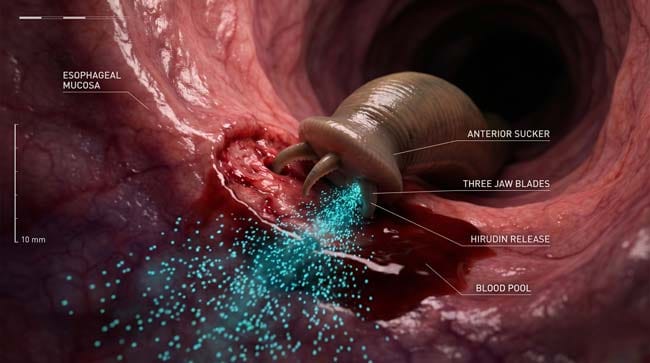
My 3D Discovery: Rendering the “Living Parasite” System
When I was setting up the hirudin molecular model for this simulation, the most striking realization was that leeches don’t just bite — they inject a complete biochemical cocktail designed to keep blood flowing indefinitely.
In the 3D viewport, I rendered the leech’s salivary glands as glowing orange reservoirs continuously pumping hirudin particles into the wound site. The anticoagulant effect is visually dramatic: blood at the attachment site refuses to clot, forming a persistent pool that the leech slowly consumes.
3D Observation: The anterior sucker mechanics surprised me. I expected simple suction. Instead, the leech uses a three-jawed rasping mechanism — shown in close-up as three blade-like structures oscillating back and forth while anchoring the sucker. The posterior sucker serves as a stable base for feeding. In the animation, you can see the leech’s body expand visibly as it ingests blood (up to 10× its body weight).
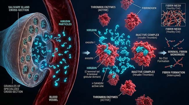
Stage 1: The Anticoagulant Cocktail — Hirudin’s Power
Leech saliva contains over 20 bioactive compounds. The primary one is hirudin — a polypeptide that irreversibly binds to thrombin, neutralizing its clot-forming ability completely.
Why This Matters Internally:
If a leech attaches to your esophageal or gastric mucosa, hirudin diffuses into surrounding tissue. Unlike a surface bite where pressure can stop bleeding, internal attachments face no external pressure. The anticoagulant effect combined with continuous feeding = ongoing blood loss that can become significant.
Hirudin Mechanism in 3D:
In our molecular simulation, I rendered hirudin molecules (blue-green particles) seeking out thrombin proteins (reddish enzymes) and binding permanently. Once bound, thrombin cannot convert fibrinogen to fibrin — the essential first step in clot formation. In the animation, the blood pool at the wound site remains liquid and flowing for the entire feeding duration (typically 20 minutes to several hours).
Other Salivary Compounds:
| Compound | 3D Color | Function | Internal Risk |
|---|---|---|---|
| Hirudin | Blue-green | Potent anticoagulant | Prevents clotting, prolonged bleeding |
| Destabilase | Yellow | Dissolves existing clots | Reactive bleeding even after leech detaches |
| Hyaluronidase | Purple | “Spreading factor” | Increases tissue permeability, enhances penetration |
| Acetylcholinesterase | White | Inhibits nerve signals | May mask pain, allowing attachment to go unnoticed |
| Aeromonas bacteria | Red | Pathogenic | Can cause cellulitis, sepsis in immunocompromised |
According to the CDC, Aeromonas infections from leech bites are rare but documented, particularly in tropical regions where leech species carry more pathogenic strains. CDC: Leech-Related Infections
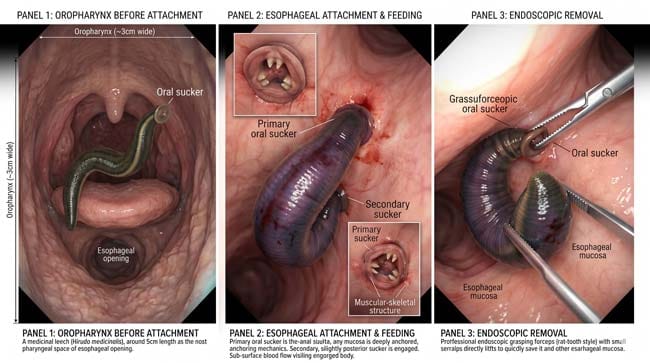
Stage 2: Attachment Sites — Esophagus vs. Stomach
Where a Leech Can Attach Internally:
1. Oropharynx / Throat (Most common if swallowed)
- Mucosa: non-keratinized stratified squamous epithelium
- Blood supply: rich, from lingual and facial arteries
- Risk: airway obstruction if large, difficulty swallowing, drooling
- Visibility: often visible on exam, may be removable with forceps
2. Esophagus
- Mucosa: similar stratified squamous
- Blood supply: inferior thyroid artery, esophageal branches
- Risk: less common attachment site but possible; can cause odynophagia (painful swallowing), hematemesis if bleeding
- 3D Visualization: esophagus shown with muscular peristalsis waves while leech anchors via anterior/posterior suckers
3. Stomach (Least likely)
- Mucosa: simple columnar epithelium with gastric pits
- Environment: pH 1.5–3.5 (strong acid)
- Risk: stomach acid is hostile to leech survival. Most leeches cannot maintain attachment long-term in acidic gastric environment.
- Exception: some species (e.g., Limnatis nilotica) have been documented in stomachs, likely swallowed and briefly survived before expelling or dying.
Attachment Mechanics (3D Breakdown):
In our anatomical model, the leech’s anterior sucker attaches to mucosa through:
- Suction vacuum created by muscular contraction
- Three jaws rasping into tissue for mechanical grip
- Secretion of adhesive compounds from salivary glands
The posterior sucker maintains attachment during feeding cycles, creating an anchor point as the leech’s body elongates to ingest blood.

Stage 3: Clinical Timeline and Medical Response
Timeline of Internal Leech Infestation:
| Hours After Swallow | Biological Event | 3D Visual | Typical Symptoms |
|---|---|---|---|
| 0–2 | Leech passes pharynx, may attach in esophagus | Leech navigating throat anatomy, seeking attachment site | Throat tickle, urge to swallow, mild discomfort |
| 2–6 | Attachment established, feeding begins | Anterior sucker anchored, hirudin particles spreading, blood pool forming | Increasing throat pain, drooling, inability to swallow saliva |
| 6–12 | Active feeding (20 min to several hours total), potential blood loss | Leech body visibly distended with ingested blood, continuous anticoagulant flow | Hemoptysis or hematemesis (coughing/vomiting blood), weakness, dizziness if significant blood loss |
| 12–24 | Leech may detach spontaneously or remain attached | Post-detachment: wound may continue bleeding due to residual hirudin | Continued bleeding, risk of secondary infection from Aeromonas |
| 24–72 | Resolution vs. complication | Wound healing vs. infection visualization | Fever, increasing pain, swollen lymph nodes = possible infection requiring antibiotics |
Medical Intervention:
- Removal: Forceps extraction with careful sucker detachment (pulling from posterior end may cause jaw tissue to remain embedded)
- Chemical deterrence: Salt, vinegar, or local anesthetic (lidocaine) can cause detachment but are not recommended for internal use
- Antibiotics: If Aeromonas infection suspected (cellulitis, erythema around wound site, fever)
- Endoscopy: For esophageal/stomach attachments, visualization and removal via endoscope
- Supportive: IV fluids if significant blood loss, blood transfusion rare but possible in extreme cases
Complications (Rare):
- Significant blood loss (<500mL usually, but larger leeches can consume more)
- Esophageal perforation (extremely rare, from prolonged attachment)
- Secondary Aeromonas hydrophila infection → cellulitis → sepsis (particularly in immunocompromised)
- Airway obstruction (if large leech in oropharynx)
According to a case series published in the Journal of Laryngology & Otology, internal leech attachments in the upper airway are uncommon (<50 documented cases annually worldwide) but can be successfully treated with prompt removal and antibiotic prophylaxis.
FAQ: What Happens If You Swallow a Leech?
Q1: Can a leech survive in my stomach acid? Most leeches cannot survive prolonged exposure to gastric acid (pH 1.5–3.5). If the leech passes into the stomach, it will likely die and be expelled naturally. The real danger is attachment in the esophagus or throat before reaching the stomach.
Q2: How much blood can a leech actually drink? A single medicinal leech (Hirudo medicinalis) can consume 5–15 mL of blood in a single feeding (up to 10× its body weight). That’s roughly 1–3 teaspoons. While not typically life-threatening, internal attachment can cause prolonged oozing due to hirudin anticoagulant effects.
Q3: Will I know if a leech is attached inside me? Yes. Symptoms usually develop within hours: persistent throat pain, feeling of a foreign body, difficulty swallowing, drooling, and potentially blood in saliva or vomit. The leech’s presence will be noticeable.
Q4: Should I try to make myself vomit to expel it? No. Vomiting can cause the leech to attach higher in the esophagus or even the pharynx. Seek medical evaluation instead. An ENT specialist can visualize the area with a scope and remove the leech safely.
Q5: Do leeches carry diseases? Yes. Leeches (especially wild freshwater species) commonly carry Aeromonas hydrophila bacteria in their gut. This can cause cellulitis, abscess formation, or sepsis if it enters through the bite wound. Antibiotics (typically trimethoprim-sulfamethoxazole or fluoroquinolones) are often given prophylactically after leech removal.
Q6: Are there documented cases of people dying from swallowed leeches? Extremely rare. Most documented cases involve complications like massive hemorrhage (usually from multiple large leeches) or secondary infection in vulnerable individuals. A few case reports exist of children with heavy leech infestations experiencing significant blood loss.
Q7: Why don’t leeches just dissolve in stomach acid? Annelid worms have protective mucus layers that provide temporary acid resistance. However, stomach acid eventually penetrates and kills them. The critical period is the first 30–60 minutes before reaching the stomach — if attachment occurs in the esophagus during that window, the leech may survive and feed for hours before either detaching or being dislodged.
Conclusion: Nature’s Blood-Sucking Machine Meets Human Biology
Leeches are evolutionary masterpieces of blood harvesting — with anticoagulants, mechanical suckers, and digestive systems optimized for liquid meals. When that mechanism operates inside your body rather than on your skin, the consequences shift from localized bleeding to potential internal complications.
The good news: internal leech infestations are rare, symptoms are usually noticeable, and medical removal is straightforward. The combination of esophagus anatomy, potential stomach acid exposure, and modern medical care means swallowed leeches are usually a brief, unpleasant episode — not a chronic parasitic infection.
In 3D, visualizing hirudin molecules flooding an attachment site and watching the anticoagulant cascade unfold helps explain why even a brief attachment can cause prolonged bleeding. It’s a reminder that evolutionary adaptations work perfectly in their natural context — whether that’s a leech on a riverbank or (unfortunately) a leech in your throat.
Further Study & External Research
- CDC — Leech-Related Infections and Aeromonas
- WHO — Waterborne Parasitic Diseases
- Journal of Laryngology & Otology — Internal Leech Infestation Case Reports
3D Simulation Specs & Observations
| 3D Component | Technical Visual Setting | Observation from Viewport |
|---|---|---|
| Framerate | 120 FPS High-Speed | Captured hirudin molecule diffusion and sucker attachment mechanics |
| Material/Shader | Subsurface Scattering (SSS) | Simulating translucency of mucosal tissue and leech body wall |
| Physics Engine | Volumetric Particle System + Soft Body Dynamics | Visualized hirudin anticoagulant particles, blood flow disruption, leech body deformation during feeding |
| Goal | Educational / Science Visualization | Research-referenced 3D breakdown of leech biology and internal attachment consequences |
Read more on Swallowed a Leech? The Survival Story of a Blood-Sucking Worm Inside You
Pingback: Swallowed a Leech? The 3D Survival Guide Inside You