
The Creator’s Note & Disclaimer: As a 3D artist at WhatIfBody3D, I rendered this scenario at 120 FPS. Our models explore foot nerve density pain science — visualizing the plantar mechanoreceptor landscape, the neurological basis of foot sensitivity, how the foot’s extraordinary nerve density serves balance and locomotion, and why the same biology that makes walking precise makes stepping on small objects so acutely painful. This visualization is part of our “What If” series and is for educational and informational purposes only, as stated in our About Page.
Quick Answer: What Is the Science Behind Foot Nerve Density and Pain? (The Atomic Answer)
Foot nerve density pain science explains one of the most counterintuitive facts in human neurology — the foot, which carries the body’s entire weight and is subject to constant mechanical stress, is simultaneously one of the most exquisitely sensitive sensory organs in the body.
- The Density: The sole of the foot contains approximately 200,000 nerve endings per square inch — a concentration comparable to the fingertips and surpassed only by the lips and tongue among non-specialized body surfaces.
- The Purpose: This extreme sensitivity is not incidental — it is the neural infrastructure for the continuous, real-time ground assessment that makes bipedal balance possible. Every step involves the foot’s mechanoreceptors sending thousands of signals per second to the brain and spinal cord.
- The Paradox: The same nerve density that enables barefoot walking over complex natural terrain — detecting slope, texture, compliance, and temperature — makes the foot extraordinarily vulnerable to small, hard objects like LEGO bricks.
- The Clinical Significance: Foot nerve density is clinically measurable — and its loss (from diabetes, peripheral neuropathy, or age) produces both reduced pain sensitivity to injury and severely impaired balance — demonstrating that the pain function and the balance function are served by the same neural infrastructure.
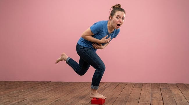
My 3D Discovery: Rendering the “Neural Landscape”
When I was building the plantar mechanoreceptor map for this simulation, the most visually stunning element was the sheer density of sensory structures visible at cellular resolution. The foot sole shown as a landscape of extraordinary biological complexity — not simply skin, fat, and bone, but an intricately organized sensory organ with different receptor types occupying specific spatial arrangements optimized for different aspects of ground contact information.
In the 3D viewport, the Meissner’s corpuscles shown arranged in precise patterns at the dermal papillae — their superficial position shown optimizing their detection of texture and light contact. The Pacinian corpuscles shown positioned deeper — their large size and layered structure shown enabling detection of vibration and rapid pressure changes across wide areas. The free nerve endings shown permeating every layer — the omnipresent pain and temperature surveillance system.
3D Observation: The most scientifically compelling visualization in this simulation is the real-time receptor activity during a single barefoot step on natural ground. I rendered the complete step — heel strike, midstance, toe-off — tracking receptor activation patterns across the entire plantar surface simultaneously. The result shown as a continuously changing, beautifully organized pattern of activation — hundreds of mechanoreceptors shown firing in precisely timed sequences as the ground contact pattern shifts throughout the step. The foot shown as a real-time tactile imaging system, providing the brain with a continuous, high-resolution map of the ground surface it is navigating.
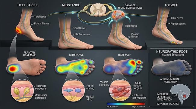
Stage 1: The Complete Plantar Sensory Landscape
The Four Mechanoreceptor Types — Specialized Functions:
Meissner’s Corpuscles — The Texture Detectors
In our 3D cellular model, Meissner’s corpuscles shown as elongated, encapsulated structures in the dermal papillae — the finger-like projections of dermis extending into the epidermis. Each corpuscle shown as approximately 40–100 micrometers long — visible at the microscopic level as small oval structures.
Functional specialization:
- Rapidly adapting — shown responding to the onset and offset of contact but not to sustained pressure
- Small receptive field — shown detecting stimuli within approximately 2–5mm of their location
- Optimal stimulus: texture changes, edges, and motion across the skin surface
- Frequency response: 10–50 Hz — optimized for detecting texture during walking
Why high density in the foot: Each step shown involving the plantar skin sliding slightly over the ground surface as weight is transferred — this sliding motion shown as optimally detected by Meissner’s corpuscles, providing real-time texture and slip information.
Merkel’s Discs — The Pressure Maps
Shown as disc-shaped receptor complexes at the basal epidermis — each shown as a specialized Merkel cell paired with an expanded nerve terminal. Present at approximately 20–30 per cm² in plantar skin.
Functional specialization:
- Slowly adapting Type I — shown maintaining firing rate throughout sustained contact
- Very small receptive field — approximately 2–3mm — providing high spatial resolution
- Optimal stimulus: sustained pressure, fine texture, curvature
- Clinical role: shown as essential for detecting the precise distribution of plantar pressure during standing and walking
Why essential for foot function: Weight distribution during standing shown requiring continuous, precise monitoring of where pressure is concentrated across the plantar surface — Merkel’s discs shown providing this sustained pressure map in real time.
Pacinian Corpuscles — The Vibration Sensors
Shown as the largest mechanoreceptors in the body — each up to 1mm long with a characteristic layered (onion-like) structure of concentric lamellae. Located in the deep dermis and plantar fascia.
Functional specialization:
- Rapidly adapting Type II — shown responding only at stimulus onset and offset
- Very large receptive field — shown detecting vibration across many centimeters
- Optimal stimulus: vibration at 200–300 Hz — the frequency range of footsteps on different surfaces
- Information provided: surface compliance (hard vs. soft), terrain type, footstep vibrations
Why essential for foot function: The vibration produced by each footstep shown propagating through the ground and returning information about surface type — shown as barefoot walkers detecting surface differences that shoe-wearers cannot. Pacinian corpuscles shown as the receptors that allow experienced barefoot walkers to detect the difference between grass, dirt, and pavement by vibration signature.
Ruffini Endings — The Stretch Sensors
Shown as elongated, spindle-shaped endings embedded in the deep dermis — oriented parallel to the skin surface. Less prominent in the plantar surface than other receptors but critically important for:
- Skin stretch detection — shown as detecting the direction and magnitude of skin deformation during lateral forces
- Joint position contribution — shown as providing proprioceptive information about foot position
- Slowly adapting Type II — shown maintaining firing throughout sustained stretch
Free Nerve Endings — The Pain and Temperature System
Present throughout all layers of the plantar skin — the highest-density receptor type. Two subtypes shown in our 3D model:
A-delta free nerve endings (thin, lightly myelinated):
- Fast pain — sharp, immediate, well-localized
- Temperature (cold preferring)
- Response threshold: lower — shown activating at moderate mechanical stimuli
C-fiber free nerve endings (unmyelinated):
- Slow pain — burning, aching, poorly localized
- Temperature (heat preferring)
- Itch — some C-fibers specialized for itch
- Response threshold: higher — shown requiring more intense stimulation
| Receptor Type | Location | Density (plantar) | Primary Detection | Role in Walking |
|---|---|---|---|---|
| Meissner’s corpuscles | Dermal papillae | 25–50/cm² | Texture, slip, light touch | Surface texture during step |
| Merkel’s discs | Basal epidermis | 20–30/cm² | Sustained pressure, fine detail | Pressure distribution mapping |
| Pacinian corpuscles | Deep dermis/fascia | 3–5/cm² | Vibration (200–300 Hz) | Surface type, impact detection |
| Ruffini endings | Deep dermis | Variable | Skin stretch, direction | Foot position sensing |
| Free nerve endings (A-delta) | All layers | Extremely high | Sharp pain, cold | Threat detection |
| Free nerve endings (C-fiber) | All layers | Extremely high | Slow pain, heat, itch | Threat signaling |
According to the Journal of Neurophysiology, the plantar surface shows regional variation in mechanoreceptor density — with the highest density at the heel and ball of the foot (primary weight-bearing regions) and the toe pads — reflecting the specific functional demands of bipedal locomotion. JNeurophysiol: Plantar Mechanoreceptor Distribution
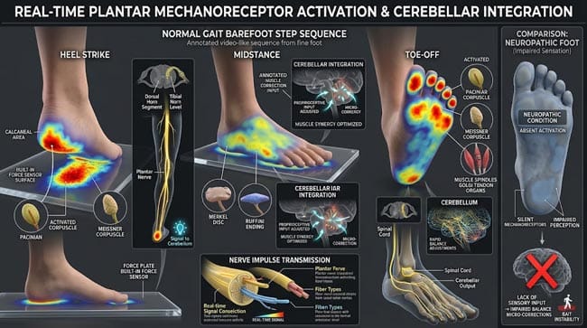
Stage 2: Why Foot Sensitivity Serves Balance — The Sensorimotor Connection
The Foot-Brain Communication System:
The foot’s mechanoreceptors shown providing three distinct types of information essential for bipedal locomotion:
Type 1 — Ground Surface Information (Exteroception)
In our 3D gait analysis model, each step shown involving the plantar mechanoreceptors providing continuous information about:
- Surface slope (via Ruffini endings detecting skin stretch asymmetry)
- Surface texture and friction (via Meissner’s corpuscles)
- Surface compliance and vibration signature (via Pacinian corpuscles)
- Pressure distribution (via Merkel’s discs)
This information shown being processed by the cerebellum and primary somatosensory cortex — shown generating the micro-adjustments in ankle, knee, and hip muscle activity that maintain balance throughout each step.
Type 2 — Foot Position Information (Proprioception)
The foot’s mechanoreceptors shown contributing to proprioception — the body’s sense of its own position — by providing:
- Information about foot contact angle at heel strike
- Detection of center of pressure shifts during stance
- Input for the vestibular system’s integration of body orientation
Type 3 — Threat Detection (Nociception)
The free nerve endings shown providing real-time threat surveillance of the plantar surface — detecting:
- Sharp objects (immediate high-pressure nociceptor activation)
- Hot surfaces (thermal nociceptors)
- Cutting or penetrating forces (polymodal nociceptors)
The Balance Demonstration — What Happens When Foot Sensitivity Is Lost:
In our 3D peripheral neuropathy model, I showed what happens to balance when plantar mechanoreceptor sensitivity is reduced:
Diabetic peripheral neuropathy: Small fiber neuropathy shown progressively reducing C-fiber and A-delta function. Then large fiber neuropathy shown affecting mechanoreceptors. The result shown as dramatically impaired balance — shown as patients with significant neuropathy shown having:
- Inability to detect ground surface characteristics
- Loss of the micro-correction system that normally maintains balance
- 2–4 times higher fall rate than age-matched controls
- 15–20 times higher risk of undetected plantar injury leading to ulceration
This balance impairment shown as having the same mechanism as the sensitivity that makes LEGO painful — the loss of both occurs together because they depend on the same sensory infrastructure.
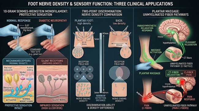
Stage 3: The Clinical Applications — From Reflexology to Neuropathy Testing
Reflexology — The Scientific Perspective:
Reflexology proposes that specific plantar zones correspond to specific organs — and that stimulation of these zones produces therapeutic effects in the corresponding organs. In our 3D model, I showed what the scientific evidence actually supports:
What is documented:
- Plantar massage shown producing generalized relaxation — shown via reduced cortisol, heart rate, and muscle tension
- Vagal stimulation shown occurring through the foot’s dense nerve supply — shown producing parasympathetic nervous system activation
- Improved local circulation shown following plantar massage — via mechanical compression of plantar vessels
What is not documented:
- No anatomical connection between specific plantar zones and specific organs
- The “organ map” claimed by reflexology shown having no correspondence to actual plantar nerve distributions or lymphatic pathways
- No peer-reviewed clinical evidence for organ-specific therapeutic effects beyond generalized relaxation
Plantar Monofilament Testing — Clinical Neuropathy Assessment:
The Semmes-Weinstein monofilament test shown as the standard clinical assessment of plantar protective sensation. A 10-gram monofilament (a thin nylon fiber) shown being applied to specific plantar sites — patients shown reporting whether they feel the touch.
In our 3D clinical model:
- Normal response: Meissner’s corpuscles and Merkel’s discs shown detecting the 10g force — protective sensation intact
- Abnormal response: Large fiber mechanoreceptors shown damaged — patient shown unable to feel the monofilament — shown indicating loss of protective sensation
Loss of protective sensation shown as the primary predictor of diabetic foot ulcer development — shown as patients shown unable to detect minor injuries, repetitive pressure, or thermal hazards that accumulate into serious wounds.
The Two-Point Discrimination Test:
Another clinical test for plantar sensitivity — measuring the minimum distance at which two simultaneous touch stimuli shown being perceived as two separate points rather than one.
- Normal plantar two-point discrimination: approximately 3–10mm — varying by region
- Fingertip two-point discrimination: approximately 2–3mm — slightly better spatial resolution
- Back two-point discrimination: approximately 40–70mm — dramatically lower density
The foot shown as having significantly better two-point discrimination than most body surfaces — shown reflecting its high mechanoreceptor density.
FAQ: Foot Nerve Density Pain Science
Q1: Why are some areas of the foot more sensitive than others? Mechanoreceptor density shown varying significantly across the plantar surface. The heel and ball of foot (metatarsal heads) shown as having the highest mechanoreceptor density — corresponding to the primary weight-bearing regions where accurate pressure information is most critical. The arch shown as having lower density — it contacts the ground only intermittently and primarily serves a shock-absorption function. The toe pads shown as having very high density — important for the push-off phase of gait and fine tactile discrimination.
Q2: Do people who walk barefoot regularly have different foot sensitivity? Yes — studies comparing habitual barefoot populations with habitual shoe-wearers show several differences. Habitual barefoot walkers shown having a thicker plantar fat pad but not necessarily reduced nerve density. Their central nervous system shown displaying better integration of plantar sensory signals — shown as more efficient balance responses to plantar stimuli. Interestingly, habitual barefoot walkers shown as having lower peak plantar pressure during walking due to different gait mechanics — shown adopting a mid-foot or fore-foot strike pattern rather than the heel-strike pattern of shoe-wearers.
Q3: Why does foot massage feel good if the foot is so sensitive to pain? The same nerve density that makes the foot responsive to painful stimuli shown also making it highly responsive to pleasant stimuli. Gentle massage shown activating C-tactile afferents (CT fibers) — a specific subset of unmyelinated fibers shown responding preferentially to slow, gentle stroking at approximately 3–5 cm/s — shown producing pleasant, rewarding sensations. These CT fibers shown projecting to the insula and anterior cingulate cortex — shown producing the relaxation and reward associated with massage. The same fiber type responsible for pleasant touch shown being distinct from the pain C-fibers — they share the same nerve but shown having different central projections and different stimulus preferences.
Q4: Can you train yourself to feel less pain when stepping on objects? Habituation to specific plantar stimuli shown occurring to some extent — repeated gentle exposure to particular textures shown producing reduced response to those stimuli through descending inhibition. However, the acute pain response to unexpected sharp plantar stimuli shown being highly resistant to habituation — because this represents a genuine threat that the nervous system is designed to respond to consistently. People who regularly walk barefoot shown reporting reduced sensitivity to minor plantar discomfort — but shown still responding fully to sharp unexpected stimuli.
Q5: Is foot ticklishness related to the same nerve density? Yes — ticklishness on the foot shown arising from the same dense mechanoreceptor population, particularly Meissner’s corpuscles detecting light, moving touch. The tickle response shown involving the same sensory pathways but processed differently in the brain — shown as light touch in an unpredictable pattern from another person shown activating both the somatosensory cortex and the anterior cingulate cortex (emotional processing). Self-tickling shown being largely ineffective because the cerebellum shown predicting self-generated touch — shown canceling the “surprise” component that generates the tickle response. The same neural infrastructure shown producing ticklishness, sensitive touch, pain from small objects, and balance information — a remarkable multi-functional sensory system.
Conclusion: The Most Functional Sensitivity in Biology
The plantar foot’s extreme nerve density is not an accident of anatomy — it is the neurological foundation of one of evolution’s greatest achievements: stable, efficient, adaptive bipedal locomotion. Every step involves the foot acting as a sophisticated sensory organ — providing the brain with real-time, high-resolution information about the ground surface that enables the continuous micro-adjustments maintaining upright balance.
In 3D, rendering the plantar mechanoreceptor landscape at cellular resolution — watching the different receptor types activate in beautifully organized patterns during a single barefoot step — transforms the foot from a structural support structure into the remarkable sensory organ it actually is.
The same biology that makes the foot able to detect a single grain of sand is the biology that makes stepping on a LEGO brick an experience nobody forgets.
Further Study & External Research
- Journal of Neurophysiology — Plantar Mechanoreceptor Distribution
- NIH — Diabetic Peripheral Neuropathy and Balance
3D Simulation Specs & Observations
| 3D Component | Technical Visual Setting | Observation from Viewport |
|---|---|---|
| Framerate | 120 FPS High-Speed | Captured mechanoreceptor activation patterns and gait sensory dynamics |
| Material/Shader | Subsurface Scattering (SSS) | Simulating plantar skin tissue layers and receptor structure visualization |
| Physics Engine | Volumetric Particle System | Visualized receptor activation patterns, signal propagation, balance correction |
| Goal | Educational / Science Visualization | Research-referenced 3D breakdown of plantar nerve density science and function |
Read more on
Read more on Why Does Stepping on a LEGO Hurt So Much?
Pingback: Why Does Stepping on a LEGO Hurt So Much? The 3D Science