
The Creator’s Note & Disclaimer: As a 3D artist at WhatIfBody3D, I rendered this scenario at 120 FPS. Our models explore why you should never pluck nose hairs — visualizing nasal hair’s filtration and immune functions, the wound created by follicle trauma, bacterial pathways through the facial danger triangle, and the infection cascade that can follow nose hair plucking in the nasal vestibule. This visualization is part of our “What If” series and is for educational and informational purposes only, as stated in our About Page.
Quick Answer: Why Should You Never Pluck Nose Hairs? (The Atomic Answer)
Why you should never pluck nose hairs comes down to what nose hairs actually do, what plucking destroys, and where the resulting wound is located.
- The Function: Nasal hairs (vibrissae) are not cosmetic waste — they are the first line of the respiratory defense system, filtering particles larger than 10 micrometers from inhaled air before they reach the lower respiratory tract.
- The Wound: Plucking a nose hair does not simply remove the hair — it tears the hair from its follicle, leaving an open wound in the nasal vestibule. This wound is surrounded by warm, moist tissue heavily colonized by bacteria — including Staphylococcus aureus.
- The Location: The nasal vestibule sits within the facial danger triangle — the region whose veins drain directly into the cavernous sinus inside the skull without valves to prevent retrograde bacterial flow.
- The Risk: A contaminated wound in this specific location creates a direct bacterial pathway to the cavernous sinus — the same pathway that makes pimple-popping in the danger triangle potentially life-threatening.
My 3D Discovery: Rendering the “Microscopic Wound”
When I was building the follicle trauma model for this simulation, the most visually striking element was the scale of damage created by nose hair plucking. At normal scale, plucking a nose hair appears to be a trivial cosmetic act. At the microscopic level, it creates a wound that would be clinically significant anywhere else on the body.
In the 3D viewport, I rendered the nose hair follicle at cellular resolution — the hair shaft shown embedded in a follicle unit extending into the dermis of the nasal vestibule, surrounded by sebaceous glands, blood capillaries, and sensory nerve fibers. When the hair is pulled, the follicle shown tearing — not cleanly, but traumatically — with the follicle wall shown rupturing, capillaries shown breaking, and the dermis shown exposed to the environment.
3D Observation: The most alarming visualization in this simulation is the bacterial access sequence. The nasal vestibule shown as one of the most bacterially contaminated surfaces in the body — its warm, moist surface colonized by dense populations of Staphylococcus aureus and other skin commensals. Immediately after follicle rupture, the bacteria shown present on the surrounding vestibule skin shown accessing the open follicle wound — shown as a sudden invasion of the previously protected follicle space. The warm, nutrient-rich environment of the torn follicle shown as ideal for rapid bacterial multiplication.
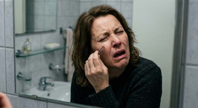
Stage 1: What Nose Hairs Actually Do — The Biology of Vibrissae
The Nasal Hair Defense System:
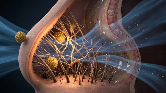
The vibrissae — coarse nasal hairs in the anterior nasal vestibule — are the visible component of the nose’s particle filtration system. In our 3D airflow model, I rendered the complete filtration mechanism:
Particle Size Filtering: Vibrissae shown as thick, irregularly arranged hairs creating a physical mesh in the nostril entrance. Airborne particles shown approaching in the inhaled airstream:
- Large particles (>10 micrometers): Pollen grains, large dust particles, insects — shown being intercepted by vibrissae before entering the nasal cavity
- Medium particles (2–10 micrometers): Shown passing through vibrissae but being captured by the mucus-coated turbinates deeper in the nasal cavity
- Fine particles (<2 micrometers): Bacteria, viruses, fine dust — shown traveling to the mucociliary system for removal
The Humidity and Temperature Functions:
Beyond filtration, nasal hairs shown contributing to:
- Air humidification: The hair surface area increasing contact time between inhaled air and the moist vestibule mucosa
- Temperature conditioning: Shown as air being warmed before reaching the more sensitive lower respiratory tract
The Microbiome Habitat:
Nasal hairs also serve as a microbiome anchor — shown hosting a commensal bacterial community that occupies the vestibule surface and competitively excludes more pathogenic species. The vibrissae shown as covered with a biofilm of commensal bacteria — primarily Staphylococcus epidermidis and Corynebacterium species — that maintain the vestibule’s ecological balance.
What Plucking Destroys:
In our comparative model, I showed the nasal filtration efficiency with and without vibrissae:
| Particle Size | With Full Vibrissae | After Significant Plucking | Consequence |
|---|---|---|---|
| >10 micrometers | 90%+ filtered | 60–70% filtered | More pollen and large particles reach turbinates |
| 5–10 micrometers | Partially filtered | Minimal filtering | Increased lower respiratory tract exposure |
| Dust and debris | Substantial trapping | Reduced trapping | More particles reach sinuses |
| Microbiome habitat | Maintained | Disrupted | Commensal community destabilized |
According to the American Academy of Otolaryngology, nasal hair density has been inversely correlated with the severity of seasonal allergic rhinitis in some studies — with denser vibrissae providing measurably better pollen filtration and reduced allergen penetration to the mucosal surface. AAO: Nasal Hair and Respiratory Defense
Stage 2: The Follicle Wound — What Plucking Actually Creates
The Anatomy of Nasal Hair Follicles:
Nasal vibrissae are housed in follicle units in the nasal vestibule — the anterior portion of the nasal cavity lined with skin (rather than the mucosal epithelium of the deeper nasal cavity).
In our 3D follicle model, I rendered the nasal vibrissa follicle:
- Hair shaft: Thick, coarse compared to body hair
- Follicle wall: Extending 3–5mm into the nasal vestibule dermis
- Sebaceous gland: Attached at the isthmus — producing sebum that lubricates the hair and contributes to the vestibule’s antimicrobial environment
- Blood supply: Rich capillary network surrounding the follicle — the nasal vestibule is highly vascular
- Nerve supply: Dense sensory nerve endings — explaining why nose hair plucking is painful
The Plucking Trauma:
When a nose hair is plucked, several simultaneous injuries occur in the 3D simulation:
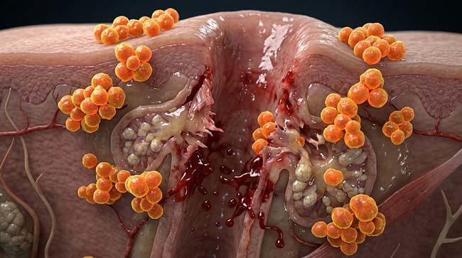
Follicle wall rupture: The hair shaft shown being pulled forcibly — the follicle wall shown unable to release the hair cleanly. At the isthmus level — the narrowest point — shown tearing. The follicle shown pulling partially inside-out as the hair exits — shown rupturing the follicle wall and exposing the underlying dermis.
Capillary rupture: The perifollicular capillaries shown tearing with the follicle trauma — producing the characteristic small bleeding that often follows nose hair plucking. In the simulation, blood shown pooling at the follicle base — providing both a nutrient-rich substrate for bacterial growth and a direct conduit to the bloodstream for anything infecting the wound.
Sebaceous gland trauma: The sebaceous gland shown partially disrupted — its contents shown released into the surrounding tissue. Sebum released into the dermis shown shown triggering a mild inflammatory response — contributing to the post-plucking tenderness.
The Resulting Wound:
In our 3D wound assessment model, I categorized the plucking wound:
- Type: Open traumatic follicle wound
- Depth: 3–5mm into the nasal vestibule dermis
- Bacterial environment: Immediately surrounded by nasal vestibule microbiota including S. aureus
- Blood supply: Highly vascular — ideal bacterial growth conditions
- Location: Within the facial danger triangle’s vascular drainage zone
- Natural protection: Lost — the protective hair and follicle sebum barrier is gone
The Bacterial Colonization Sequence:
In our 3D infection timeline, I showed what happens to the plucking wound in an unsterile environment (the nostril):
Immediately (0–5 minutes): Bacteria shown on the surrounding vestibule skin accessing the open follicle wound — S. aureus shown as the primary early colonizer, attracted by the blood and serum at the wound base.
Hours 1–6: Bacterial multiplication within the follicle space shown — the warm, moist, nutrient-rich environment shown supporting rapid replication. Early inflammatory response shown beginning — neutrophils shown arriving at the wound site.
Hours 6–24: Two possible pathways diverge:
- Resolution: Immune response successfully contains the bacteria — shown as neutrophils eliminating the bacterial colony, wound beginning to heal
- Folliculitis: Bacteria overcome the local immune response — shown as a pustule forming at the follicle site — the beginning of nasal vestibulitis
Stage 3: The Danger Triangle Connection — Why Location Changes Everything
Why the Nasal Vestibule Is Uniquely Dangerous:
A follicle wound from plucking a body hair anywhere else would be a minor, self-limited event. The nasal vestibule’s specific anatomical location within the facial danger triangle transforms the same type of wound into a potential medical emergency.
The Vascular Pathway Review:
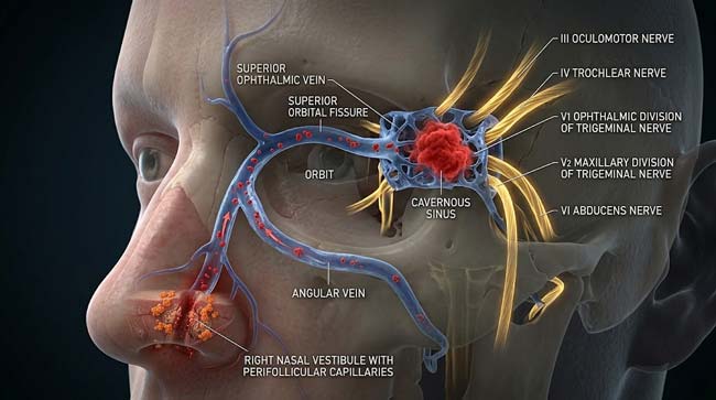
In our 3D vascular model (building on the danger triangle anatomy from the pimple-popping article), I showed the complete bacterial pathway from nasal vestibule to cavernous sinus:
Step 1: Bacteria from infected nasal vestibule follicle enter the perifollicular capillaries
Step 2: Blood from the nasal vestibule drains into the facial vein and angular vein — both within the danger triangle
Step 3: The angular vein connects to the superior ophthalmic vein — shown traveling backward through the orbit
Step 4: The superior ophthalmic vein passes through the superior orbital fissure into the skull
Step 5: Direct drainage into the cavernous sinus — the venous pool adjacent to critical cranial nerves and the brain
Step 6: Without valves to enforce directional flow — retrograde bacterial transport shown possible from the facial wound to the intracranial space
The Progression from Minor Wound to Life-Threatening:
In our 3D infection progression model, I showed three distinct outcomes depending on bacterial load and immune response:
Outcome A — Self-Limited (Most Common) Minor follicle wound shown healing without complication — immune system successfully containing the bacterial colonization. No infection develops. The risk existed but was not realized.
Outcome B — Nasal Vestibulitis Bacterial colony established in the follicle — shown developing into nasal vestibulitis (localized infection of the nasal vestibule). Presents as pain, redness, and swelling at the plucking site. Treatable with topical or oral antibiotics. Risk of progression to cavernous sinus thrombosis is low but real.
Outcome C — Cavernous Sinus Thrombosis (Rare but Documented) Bacteria shown entering the venous system from the infected follicle — traveling the retrograde pathway to the cavernous sinus. CST developing with characteristic proptosis, chemosis, and ophthalmoplegia. Carries approximately 30% mortality even with aggressive antibiotic and anticoagulation treatment.
| Outcome | Probability | Bacterial Requirement | Treatment | Mortality |
|---|---|---|---|---|
| Self-limited healing | >90% | Low bacterial load, effective immune response | None needed | 0% |
| Nasal vestibulitis | 5–10% | Moderate bacterial colonization | Antibiotics | <1% |
| Cavernous sinus thrombosis | <0.1% but documented | High bacterial load + retrograde venous seeding | Emergency treatment | ~30% |
The Preventable Nature of the Risk:
The key insight shown in our 3D comparison model: the risk from nasal vestibulitis to cavernous sinus thrombosis is not inevitable — it depends on whether an infection is allowed to establish and spread. The infection that develops in a plucked follicle is:
- Preventable by not plucking and using a trimmer instead
- Treatable if recognized early (nasal vestibulitis)
- Catastrophic if allowed to progress unrecognized to CST
According to the New England Journal of Medicine, case reports of cavernous sinus thrombosis following nose hair plucking have been documented across multiple medical centers globally — with the consistent finding that the infection arose at the plucking wound site and propagated through the facial danger triangle venous system. NEJM: Cavernous Sinus Thrombosis — Case Series
FAQ: Why You Should Never Pluck Nose Hairs
Q1: Is it safe to use a nose hair trimmer instead of plucking? Yes — completely. A trimmer cuts the visible portion of the nose hair without disturbing the follicle or the underlying skin. No wound is created. No bacterial access to the dermis occurs. No danger triangle risk is activated. Trimmers are the universally recommended alternative to plucking — they achieve the cosmetic goal of reducing visible nasal hair without any of the infection risks associated with follicle disruption.
Q2: What about nose hair waxing — is that safer than plucking? No — waxing is equally or more dangerous than plucking. Waxing removes multiple nose hairs simultaneously, each from its follicle, creating multiple simultaneous follicle wounds across the nasal vestibule. The simultaneous trauma shown in the simulation as dramatically increasing total wound area, bacterial access, and cumulative infection risk. Some dermatologists and ENT specialists consider nasal waxing more dangerous than individual plucking precisely because of the scale of simultaneous follicle disruption.
Q3: How do I know if a plucked nose hair has become infected? Signs of nasal vestibulitis following plucking include: increasing pain at the plucking site beyond the immediate post-plucking soreness, visible redness or swelling inside the nostril, formation of a pustule (whitehead) at the follicle site, and warmth of the overlying skin. These symptoms warrant medical evaluation — do not squeeze or manipulate the infected area. Antibiotics (topical mupirocin or oral antibiotics depending on severity) effectively treat early nasal vestibulitis before progression.
Q4: Are some people at higher risk from nose hair plucking than others? Yes. People with diabetes, immune suppression, chronic steroid use, or HIV are at significantly higher risk because their immune systems cannot as effectively contain the initial bacterial colonization. People who are known carriers of Staphylococcus aureus (particularly MRSA) in their nasal passages face higher risk because their colonizing bacteria are more virulent. People who pluck in unclean conditions (public bathrooms, unwashed hands) introduce more bacteria to the wound site.
Q5: What is the safest way to manage nose hair for cosmetic purposes? A quality nose hair trimmer with rounded safety tips is the gold standard. Electric rotary trimmers designed specifically for the nasal vestibule trim only the visible external portion of the hair without entering deeply enough to create wound risk. For in-salon grooming, a trained aesthetician using proper technique and sterile implements reduces risk significantly compared to home plucking. The universal recommendation from dermatologists and ENT specialists is to trim, never pluck.
Conclusion: The Most Dangerous Grooming Habit You Never Thought About
Plucking nose hairs ranks among the most underappreciated risk behaviors in routine grooming — not because the risk is high (the vast majority of plucking events cause only brief pain and minor, self-healing wounds), but because the anatomy of the nasal vestibule means that when things go wrong, they go wrong in proximity to a direct intracranial venous pathway.
In 3D, rendering the wound left by plucking — the torn follicle wall, the exposed capillaries, the immediate bacterial access from the surrounding vestibule microbiota — and then showing the vascular pathway from that wound to the cavernous sinus makes the anatomical basis of the risk immediately clear.
The risk is real. The alternative — a trimmer — is available, inexpensive, and eliminates the risk entirely.
Use a trimmer. Keep the vibrissae. Let them do their job.
Further Study & External Research
3D Simulation Specs & Observations
| 3D Component | Technical Visual Setting | Observation from Viewport |
|---|---|---|
| Framerate | 120 FPS High-Speed | Captured follicle trauma mechanics and bacterial invasion dynamics |
| Material/Shader | Subsurface Scattering (SSS) | Simulating nasal vestibule tissue and follicle wall translucency |
| Physics Engine | Soft Body Dynamics + Particle System | Visualized follicle rupture, capillary trauma, bacterial access sequence |
| Goal | Educational / Science Visualization | Research-referenced 3D breakdown of nose hair plucking infection risk |
Read more on
Pingback: Nose Hair Plucking Brain Infection: The 3D Science Guide